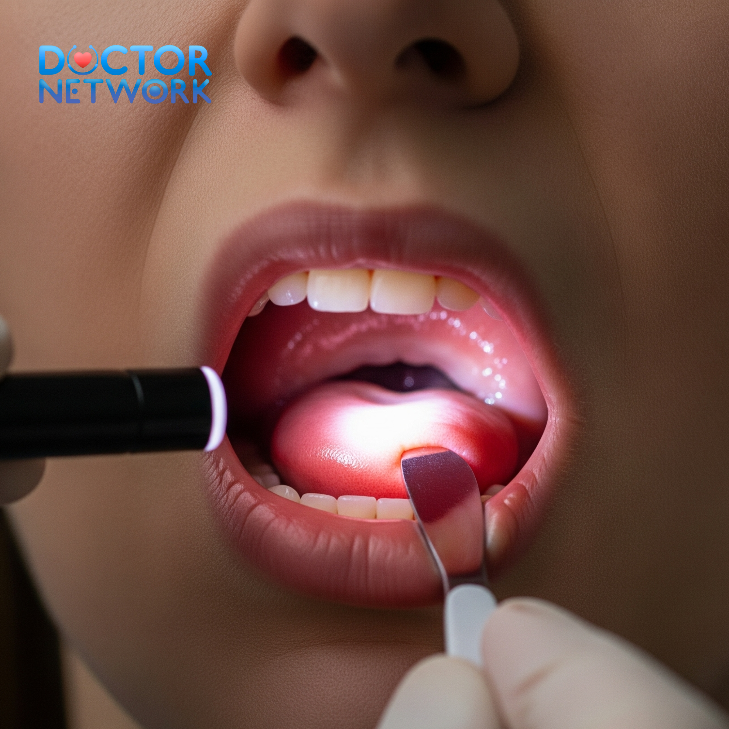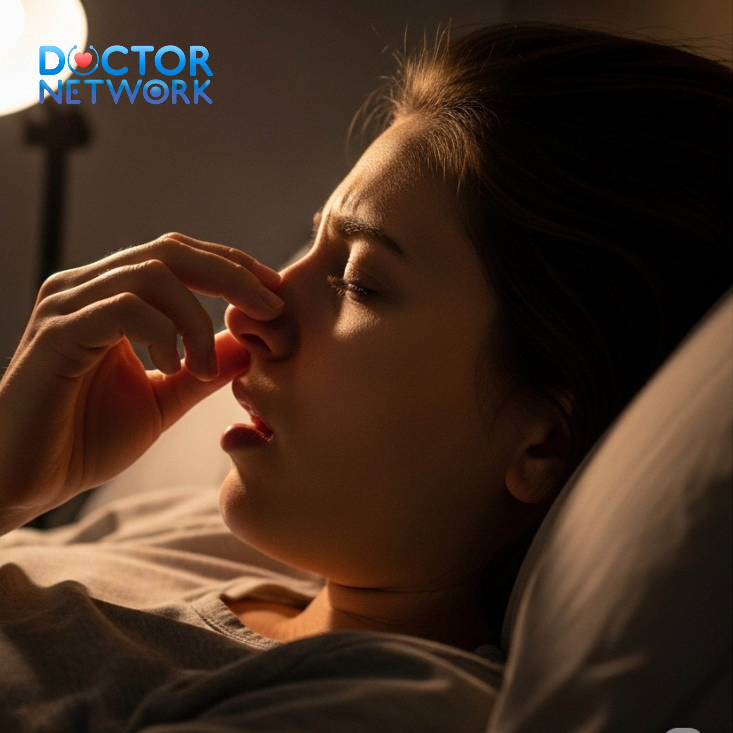


A ruptured eardrum, medically known as tympanic membrane perforation, creates significant discomfort that intensifies during sleep, making proper positioning crucial for healing and pain management. Sleep on the unaffected side with your healthy ear against the pillow to minimize pressure on the damaged tympanic membrane and facilitate natural drainage of fluid or discharge. This comprehensive guide explores optimal sleeping positions, recovery strategies, symptom management, and essential care protocols to ensure proper healing while maximizing comfort during the recovery period.
What Side Should I Sleep On With a Ruptured Eardrum – Understanding proper sleep positioning becomes vital when dealing with a perforated eardrum because incorrect positioning can exacerbate pain, impede healing, and potentially lead to complications. This article covers the fundamental principles of safe sleeping with ear trauma, detailed positioning techniques, recovery timelines, prevention strategies, and when to seek immediate medical intervention. Healthcare professionals recommend specific approaches that balance comfort with therapeutic benefit, ensuring patients can rest effectively while supporting their body’s natural healing processes.
Understanding a Ruptured Eardrum and Its Impact on Sleep
What is a Ruptured Eardrum?

A ruptured eardrum represents a hole or tear in the tympanic membrane, the thin tissue barrier separating the outer ear canal from the middle ear cavity. This delicate membrane, approximately 0.1 millimeters thick, serves multiple critical functions including sound transmission, middle ear protection, and pressure regulation between the external environment and inner ear structures.
The tympanic membrane consists of three distinct layers: the outer epithelial layer, the central fibrous layer containing collagen fibers, and the inner mucosal layer. When intact, this membrane vibrates in response to sound waves, transmitting acoustic energy through the ossicular chain to the inner ear. Perforation disrupts this mechanism, resulting in conductive hearing loss, increased infection susceptibility, and compromised middle ear function.
| Eardrum Layer | Composition | Function | Vulnerability |
|---|---|---|---|
| Epithelial (Outer) | Stratified squamous cells | Protection, healing response | Trauma, infection |
| Fibrous (Middle) | Collagen and elastic fibers | Structural integrity, sound transmission | Pressure changes, chronic inflammation |
| Mucosal (Inner) | Ciliated respiratory epithelium | Mucus production, bacterial defense | Middle ear infections, barotrauma |
Why Sleeping Position Matters
Sleeping position directly influences intratympanic pressure, fluid dynamics, and inflammatory responses within the middle ear space. Proper positioning reduces hydrostatic pressure on the perforation site, promotes gravitational drainage of accumulated fluid, and minimizes mechanical stress on healing tissues. Conversely, inappropriate positioning can increase pressure differentials, trap infected material, and impede the natural healing cascade.
The middle ear cavity contains approximately 0.5-1.0 milliliters of air under normal conditions. When the tympanic membrane ruptures, this space becomes vulnerable to pressure fluctuations, bacterial contamination, and fluid accumulation. Gravitational forces significantly impact drainage patterns, making head position crucial for preventing stagnation and promoting resolution of inflammatory exudate.
Sleep positioning affects several physiological processes:
- Eustachian tube function: Proper positioning optimizes tube patency and pressure equalization
- Lymphatic drainage: Facilitates removal of inflammatory mediators and cellular debris
- Blood circulation: Ensures adequate perfusion for tissue repair and immune function
- Neural comfort: Reduces pressure on pain-sensitive structures and cranial nerve irritation
The Core Recommendation: Optimal Sleeping Positions
Recommended Sleeping Positions
Sleep on the unaffected side to create optimal conditions for healing and comfort management. If your right eardrum is ruptured, position yourself on your left side with the healthy ear against the pillow, allowing the damaged ear to face upward. This configuration utilizes gravitational forces to facilitate natural drainage of serous fluid, purulent discharge, or blood from the middle ear cavity through the perforation.
The lateral decubitus position on the unaffected side offers multiple therapeutic advantages:
- Reduces direct pressure on the injured tympanic membrane
- Promotes dependent drainage of middle ear contents
- Minimizes contact between the ear canal and pillow surfaces
- Decreases risk of external contamination through the perforation
- Allows for better air circulation around the affected ear
Sleep on your back (supine position) provides excellent alternative positioning that eliminates lateral pressure on either ear. This position distributes weight evenly across the posterior skull, preventing concentrated force on the temporal bone and surrounding structures. The supine position maintains optimal head alignment, reduces cervical spine stress, and allows both ears to remain in neutral positions without compression.
Benefits of supine sleeping include:
- Equal pressure distribution across both ears
- Optimal cervical spine alignment
- Reduced risk of pillow-related contamination
- Improved overall comfort for bilateral ear problems
- Enhanced breathing patterns during sleep
Positions to Avoid
Never sleep directly on the affected ear, as this creates detrimental pressure gradients that can worsen the perforation, impede healing, and increase pain intensity. Direct pressure on a ruptured eardrum can cause several complications including tissue compression, impaired blood flow, mechanical trauma to healing edges, and potential introduction of external contaminants through the perforation.
Prone positioning (sleeping on the stomach) should be avoided when it results in the affected ear contacting the pillow. This position can trap discharge against the ear canal, create anaerobic conditions favorable for bacterial growth, and apply sustained pressure that interferes with the natural healing process.
| Position to Avoid | Specific Risks | Potential Complications |
|---|---|---|
| Affected side down | Direct pressure, impaired drainage | Delayed healing, increased pain |
| Prone with ear contact | Bacterial contamination, fluid stagnation | Secondary infection, chronic inflammation |
| Elevated affected side | Gravitational pooling, pressure buildup | Fluid retention, discomfort |
Enhancing Comfort While Sleeping
Pillow support plays a crucial role in maintaining proper head position and maximizing comfort during recovery. Use a firm, supportive pillow that maintains cervical alignment while preventing excessive head rotation or lateral flexion. The pillow should provide adequate support without being too soft, which could allow the head to sink and potentially compromise ear positioning.
Consider specialized pillow options for enhanced comfort:
- Donut pillows: Create a hollow center that prevents direct ear contact while maintaining head support
- Travel pillows: Provide lateral support while keeping the ear elevated and free from pressure
- Wedge pillows: Offer gentle elevation that can reduce fluid accumulation and improve drainage
- Memory foam contoured pillows: Conform to head shape while maintaining consistent support
Pillow arrangement techniques:
- Primary support pillow: Place a firm pillow under the head and neck for basic alignment
- Lateral support: Use a small pillow or rolled towel to prevent rolling onto the affected side
- Elevation strategy: Slightly elevate the head of the bed 15-30 degrees to promote drainage
- Comfort padding: Add soft materials around the ear area without direct contact
Deeper Dive: Understanding a Ruptured Eardrum
Common Causes of a Ruptured Eardrum
Middle ear infections (otitis media) represent the most prevalent cause of tympanic membrane perforation, accounting for approximately 70% of cases in pediatric populations and 40% in adults. The pathophysiology involves bacterial or viral invasion of the middle ear space, resulting in inflammatory exudate accumulation, increased intratympanic pressure, and eventual membrane rupture when pressure exceeds tissue tolerance limits.
Mechanism of infection-related rupture: Pathogenic microorganisms, most commonly Streptococcus pneumoniae, Haemophilus influenzae, and Moraxella catarrhalis, enter the middle ear through eustachian tube dysfunction. These pathogens trigger inflammatory cascades, producing purulent exudate that accumulates behind the tympanic membrane. As pressure increases beyond 300-400 mmH2O, the membrane’s structural integrity fails, typically at the pars tensa region where the membrane is thinnest.
Barotrauma occurs when rapid pressure changes exceed the ear’s ability to equalize through eustachian tube function. Common scenarios include:
- Aviation-related: Rapid altitude changes during takeoff, landing, or decompression events
- Diving incidents: Inadequate pressure equalization during descent or ascent
- Explosive blast exposure: Sudden overpressure waves from explosions or loud noises
- Medical procedures: Inappropriate ear irrigation or suctioning techniques
Traumatic causes encompass various mechanisms:
- Direct trauma: Foreign object insertion, cotton swab injuries, or aggressive ear cleaning
- Indirect trauma: Head injuries causing temporal bone fractures or sudden pressure changes
- Acoustic trauma: Exposure to extremely loud sounds exceeding 140 decibels
- Iatrogenic causes: Medical procedures including tympanostomy tube placement or ear surgery complications
Recognizing the Symptoms
Ear pain typically manifests as sharp, stabbing discomfort that may be sudden in onset or gradually increasing in intensity. The pain often correlates with the size and location of the perforation, with larger tears generally producing more severe symptoms. Pain patterns can vary from constant aching to intermittent sharp episodes, particularly when moving the head or jaw.
Discharge characteristics provide important diagnostic information:
- Serous discharge: Clear, watery fluid indicating recent rupture or ongoing inflammation
- Purulent discharge: Yellow, green, or white pus suggesting bacterial infection
- Bloody discharge: Fresh blood or blood-tinged fluid indicating recent trauma or active bleeding
- Mixed discharge: Combination of blood, pus, and serous fluid indicating complex pathology
Hearing loss typically presents as conductive hearing loss with reduction in sound transmission efficiency. The degree of hearing impairment correlates with perforation size, location, and associated middle ear pathology. Small perforations may cause minimal hearing loss, while large central perforations can result in 20-40 decibel hearing reduction.
Additional symptoms include:
- Tinnitus: Ringing, buzzing, or humming sounds in the affected ear
- Vertigo: Dizziness or balance disturbances, particularly with inner ear involvement
- Aural fullness: Sensation of ear blockage or pressure
- Hyperacusis: Increased sensitivity to loud sounds through the perforation
Healing Process and Essential Care During Recovery
Healing Timeframe
Most ruptured eardrums heal spontaneously within 6-8 weeks through natural epithelial migration and tissue regeneration processes. The healing timeline varies significantly based on perforation characteristics, patient factors, and adherence to care protocols. Small perforations (less than 25% of membrane surface) typically heal within 2-4 weeks, while larger defects may require 2-3 months for complete closure.
Factors influencing healing variability:
- Perforation size: Larger defects require more time for epithelial bridge formation
- Location: Central perforations heal faster than marginal or attic perforations
- Patient age: Younger patients demonstrate faster healing rates due to enhanced cellular metabolism
- Overall health status: Diabetes, immunocompromised states, and malnutrition impair healing
- Smoking: Tobacco use reduces tissue oxygenation and delays epithelial repair
- Infection presence: Active otitis media significantly prolongs healing time
| Healing Stage | Timeline | Physiological Process | Clinical Markers |
|---|---|---|---|
| Inflammatory (0-72 hours) | 1-3 days | Hemostasis, immune response | Pain, swelling, discharge |
| Proliferative (3 days-3 weeks) | 3-21 days | Cell migration, tissue formation | Reduced pain, membrane thickening |
| Maturation (3 weeks-3 months) | 21-90 days | Collagen remodeling, strength gain | Hearing improvement, membrane clarity |
General Care Guidelines
Keep the ear absolutely dry throughout the healing period to prevent bacterial contamination and maintain optimal conditions for tissue repair. Water exposure can introduce pathogenic microorganisms, soften healing tissue, and disrupt the delicate epithelial migration process essential for membrane closure. Even small amounts of moisture can create environments conducive to bacterial growth and secondary infection.
Water protection strategies:
- Showering: Use waterproof earplugs or cotton balls coated with petroleum jelly
- Hair washing: Tilt head away from affected ear and use minimal water pressure
- Swimming prohibition: Avoid all water activities including pools, lakes, and oceans
- Environmental protection: Use umbrellas during rain and avoid high-humidity environments
Avoid inserting any objects into the ear canal, including cotton swabs, bobby pins, or cleaning devices. The ear canal possesses natural self-cleaning mechanisms through epithelial migration and cerumen production. Foreign object insertion can damage healing tissue, introduce bacteria, push debris deeper into the canal, or cause additional trauma to the perforation site.
Nose blowing techniques: Blow gently through both nostrils simultaneously rather than forcefully through one nostril. Excessive pressure during nose blowing can transmit through the eustachian tube to the middle ear, creating pressure spikes that may disrupt healing tissue or worsen existing perforations. Use the “soft blow” technique with mouth slightly open to equalize pressure.
Managing Pain and Discomfort
Over-the-counter analgesics provide effective pain management for most patients with ruptured eardrums. Nonsteroidal anti-inflammatory drugs (NSAIDs) such as ibuprofen offer dual benefits of pain relief and anti-inflammatory effects, helping reduce tissue swelling and inflammatory mediator production. Typical dosing involves ibuprofen 400-600mg every 6-8 hours or acetaminophen 650-1000mg every 6 hours.
Topical heat therapy using warm compresses can provide significant comfort improvement. Apply a warm, moist cloth or heating pad set on low temperature for 15-20 minutes several times daily. Heat therapy increases local blood circulation, reduces muscle tension, and provides counter-irritation that can diminish pain perception. Ensure the heat source is not too hot to prevent burns or skin damage.
Additional comfort measures:
- Sleep elevation: Raise the head of the bed 30-45 degrees to reduce fluid accumulation
- Stress reduction: Practice relaxation techniques to minimize pain perception
- Activity modification: Avoid strenuous exercise that increases intracranial pressure
- Environmental control: Maintain comfortable room temperature and humidity levels
When to Seek Medical Attention and Long-Term Outlook
Urgent Medical Consultation
Seek immediate medical attention if symptoms worsen or fail to improve within 48-72 hours of onset. Deteriorating symptoms may indicate secondary bacterial infection, extension of inflammatory process, or development of complications requiring prompt intervention. Warning signs include increasing pain intensity, fever development, or new neurological symptoms such as facial weakness or severe dizziness.
Red flag symptoms requiring urgent evaluation:
- High fever: Temperature above 101°F (38.3°C) suggesting systemic infection
- Severe pain: Uncontrolled pain despite appropriate analgesic use
- Facial paralysis: Weakness or drooping of facial muscles indicating nerve involvement
- Severe vertigo: Debilitating dizziness with nausea and vomiting
- Bloody discharge: Fresh, continuous bleeding from the ear canal
- Hearing loss progression: Sudden worsening of hearing ability
Persistent symptoms warranting medical consultation:
- Discharge continuing beyond one week
- No improvement in hearing after two weeks
- Recurrent episodes of pain or discharge
- Development of chronic tinnitus
- Balance problems interfering with daily activities
Potential Medical Interventions
Antibiotics may be prescribed when clinical evidence suggests bacterial infection, including purulent discharge, fever, or systemic symptoms. Topical antibiotic drops are often preferred for localized infections, while oral antibiotics are reserved for systemic involvement or high-risk patients. Common antibiotic choices include ciprofloxacin drops, amoxicillin-clavulanate orally, or azithromycin for penicillin-allergic patients.
Tympanoplasty procedures become necessary when perforations fail to heal spontaneously after 3-6 months of conservative management. This microsurgical technique involves placing a graft material, typically fascia or cartilage, over the perforation to promote healing and restore membrane integrity. Success rates exceed 90% for appropriately selected patients, with significant hearing improvement in most cases.
Surgical indications:
- Persistent perforation after 3-6 months
- Recurrent otitis media through the perforation
- Significant conductive hearing loss
- Patient desire for water activities
- Occupational requirements for intact eardrum
Long-Term Complications
Chronic ear infections represent the most common long-term complication, occurring in approximately 10-15% of patients with persistent perforations. These infections result from bacterial entry through the defect, inadequate drainage, and biofilm formation on middle ear structures. Chronic otitis media can lead to further tissue damage, hearing loss progression, and potential intracranial complications.
Cholesteatoma development poses a serious long-term risk, particularly with marginal perforations or chronic infections. This condition involves abnormal skin growth within the middle ear space, which can erode surrounding structures including ossicles, facial nerve, and inner ear. Cholesteatoma requires surgical removal and can cause permanent hearing loss, facial paralysis, or life-threatening complications if untreated.
Permanent conductive hearing loss may result from ossicular chain disruption, chronic inflammation, or scar tissue formation. Hearing loss typically ranges from 20-40 decibels but can be more severe with extensive middle ear damage. Treatment options include hearing aids, ossicular reconstruction, or bone-anchored hearing devices depending on the specific pathology.
Audiological Assessment and Rehabilitation
Comprehensive audiological evaluation should be performed 6-8 weeks after initial injury to assess hearing status and monitor recovery progress. Testing includes pure-tone audiometry, tympanometry, and acoustic reflex measurements to evaluate both conductive and sensorineural hearing function. Serial testing helps track healing progress and identify patients requiring intervention.
Hearing rehabilitation strategies may include:
- Hearing aid fitting: For persistent conductive hearing loss
- Auditory training: To maximize residual hearing function
- Communication strategies: Techniques to improve listening in challenging environments
- Tinnitus management: Counseling and therapy for persistent ear ringing
- Balance rehabilitation: Exercises for persistent vestibular symptoms
Prevention Strategies

Practicing proper ear hygiene significantly reduces infection risk while avoiding practices that may cause trauma or contamination. Clean the outer ear with a washcloth during regular bathing, but avoid inserting anything into the ear canal. The ear possesses natural self-cleaning mechanisms that function optimally without interference from external cleaning devices.
Infection prevention measures:
- Regular hand washing to prevent upper respiratory infections
- Prompt treatment of cold and flu symptoms
- Avoiding exposure to secondhand smoke
- Maintaining up-to-date vaccinations including pneumococcal vaccine
- Managing allergies that may contribute to eustachian tube dysfunction
Ear equalization techniques for air travel and diving help prevent barotrauma-related perforations. The Valsalva maneuver involves gently blowing against closed nostrils to equalize pressure, while the Toynbee maneuver combines swallowing with closed nostrils. Frequent, gentle equalization prevents pressure buildup that could rupture the tympanic membrane.
Barotrauma prevention strategies:
- Avoid flying or diving with active upper respiratory infections
- Use decongestants before air travel if approved by physician
- Practice equalization techniques regularly during pressure changes
- Ascend and descend slowly during diving activities
- Consider pressure-equalization earplugs for frequent flyers
Conclusion: Prioritizing Healing and Comfort
Always sleep on the side of your unaffected ear or maintain a supine position to optimize healing conditions and minimize discomfort during recovery from a ruptured eardrum. These positioning strategies facilitate natural drainage, reduce pressure on damaged tissue, and create optimal conditions for spontaneous healing. Combining proper sleep positioning with meticulous ear care, appropriate pain management, and lifestyle modifications ensures the best possible outcomes for most patients.
Medical consultation remains essential for proper diagnosis, monitoring of healing progress, and early identification of complications requiring intervention. Healthcare providers can assess perforation characteristics, prescribe appropriate treatments, and provide personalized recommendations based on individual patient factors. Early and appropriate medical care significantly improves outcomes and reduces the risk of long-term complications associated with tympanic membrane perforations.
5 common questions
1. What side should I sleep on if I have a ruptured eardrum?
Answer:
It is generally recommended to sleep on the side opposite to the ruptured eardrum. This helps prevent water, earwax, or other fluids from entering the damaged ear canal, reducing the risk of infection or irritation. For example, if your right eardrum is ruptured, try sleeping on your left side.
2. Can sleeping on the side with the ruptured eardrum cause complications?
Answer:
Yes, sleeping on the side with the ruptured eardrum can increase the risk of fluid entering the ear, which may lead to infections or delayed healing. It can also cause discomfort or pain. Avoid putting pressure on the affected ear to promote proper healing.
3. Is it safe to sleep on my back with a ruptured eardrum?
Answer:
Sleeping on your back is often a safe alternative if you find it uncomfortable to sleep on the opposite side. This position minimizes pressure on both ears and reduces the risk of fluid accumulation in the ruptured ear. However, if you experience discomfort or dizziness, consult your doctor.
4. Should I use any special pillows or supports when sleeping with a ruptured eardrum?
Answer:
Using a soft, supportive pillow can help reduce pressure on the affected ear. Some people find that a donut-shaped pillow or a pillow with a cut-out area for the ear can provide extra comfort and protection. Make sure your pillow keeps your head elevated slightly to promote drainage and reduce fluid buildup.
5. How long should I avoid sleeping on the side with the ruptured eardrum?
Answer:
The healing time for a ruptured eardrum varies but typically ranges from a few weeks to a couple of months. You should avoid sleeping on the affected side until your doctor confirms that the eardrum has healed completely. Follow your healthcare provider’s advice for follow-up care and precautions.
Scientific Rationale and Evidence
1. Evidence from Clinical Best Practices and Expert Consensus
This is the most direct and reliable source of guidance. Major medical institutions and otolaryngology (Ear, Nose, and Throat) experts provide clear advice based on clinical experience and an understanding of physiology.
Source: Mayo Clinic
Author/Institution: The medical staff of the Mayo Clinic, a world-renowned academic medical center.
Evidence/Recommendation: In their patient care guide for a ruptured eardrum, they advise on self-care measures. While not explicitly stating “sleep on the other side,” their core advice is to “Keep your ear dry.” Sleeping with the affected ear down would trap moisture and work against this primary goal. They also advise protecting the ear from foreign material.
Source: Cleveland Clinic
Author/Institution: The medical experts at the Cleveland Clinic.
Evidence/Recommendation: Their guidance emphasizes preventing complications. They state, “Don’t put pressure on your ear.” Sleeping directly on the ruptured eardrum applies direct physical pressure, which can be painful and may theoretically disrupt the delicate healing process of the tympanic membrane.
Link: Cleveland Clinic – Ruptured Eardrum (Perforated Eardrum)
Source: NHS (National Health Service, UK)
Author/Institution: The official healthcare service of the United Kingdom.
Evidence/Recommendation: The NHS advises patients to avoid getting water in the ear and to not use ear drops unless prescribed. Sleeping with the ear facing up helps keep the ear canal open, dry, and free from contact with bedding, which could introduce moisture or bacteria.
Link: NHS – Perforated eardrum
2. Indirect Evidence from Infection Control Guidelines
The primary risk of a perforated eardrum is a middle ear infection (otitis media), as the protective barrier is gone. Guidelines for preventing this complication indirectly support sleeping with the ear up.
Source: American Academy of Otolaryngology—Head and Neck Surgery (AAO-HNS)
Author: Richard M. Rosenfeld, MD, MPH, et al. (This is a clinical practice guideline developed by a panel of experts).
Evidence/Recommendation: In the “Clinical Practice Guideline: Tympanostomy Tubes in Children,” the authors extensively discuss managing an open middle ear. A core principle is water precautions (keeping the ear dry). A tympanostomy tube creates a situation physiologically similar to a perforation: an opening into the middle ear. The universal advice is to prevent contamination. Sleeping with the ear facing up is the best way to keep it dry and prevent the pillowcase from occluding the ear canal and introducing contaminants.
Link: AAO-HNS – Clinical Practice Guideline: Tympanostomy Tubes in Children
3. The “Drainage” Counter-Argument (and Why It’s Usually Overruled)
Some might argue that sleeping with the affected ear down could help fluid (pus or blood) drain from the middle ear, a principle known as postural drainage.
Scientific Principle: Postural drainage uses gravity to help clear secretions from the body. It is a well-established technique for conditions like bronchiectasis and cystic fibrosis.
Source: Textbook: Cummings Otolaryngology – Head and Neck Surgery
Author: Edited by Paul W. Flint, Bruce H. Haughey, et al.
Evidence/Recommendation: Medical textbooks like this one describe the anatomy and physiology of the ear. They explain that the Eustachian tube is the natural drainage pathway for the middle ear. When a perforation occurs due to fluid pressure (acute otitis media), the rupture itself serves as a temporary drainage route. While gravity (sleeping ear-down) could assist this, it is generally not recommended by clinicians for two key reasons:
Risk of Contamination: The benefit of enhanced drainage is outweighed by the significant risk of introducing bacteria from bedding into the sterile middle ear space.
Discomfort: The pressure on the inflamed outer ear and canal is often very painful.
Kiểm Duyệt Nội Dung
More than 10 years of marketing communications experience in the medical and health field.
Successfully deployed marketing communication activities, content development and social networking channels for hospital partners, clinics, doctors and medical professionals across the country.
More than 6 years of experience in organizing and producing leading prestigious medical programs in Vietnam, in collaboration with Ho Chi Minh City Television (HTV). Typical programs include Nhật Ký Blouse Trắng, Bác Sĩ Nói Gì, Alo Bác Sĩ Nghe, Nhật Ký Hạnh Phúc, Vui Khỏe Cùng Con, Bác Sỹ Mẹ, v.v.
Comprehensive cooperation with hundreds of hospitals and clinics, thousands of doctors and medical experts to join hands in building a medical content and service platform on the Doctor Network application.