Why You Might Get More Sinus Infections After Septoplasty – Experiencing increased sinus infections after septoplasty can be alarming, but this temporary complication affects approximately 15-20% of patients during the initial healing period. Post-surgical inflammation, altered nasal drainage patterns, and compromised ciliary function create an environment where bacterial colonization thrives, leading to recurrent sinusitis episodes that typically resolve within 3-6 months as healing progresses.
This comprehensive analysis explores the underlying mechanisms causing post-septoplasty sinus infections, identifies risk factors that predispose patients to complications, examines prevention strategies, and provides evidence-based treatment approaches. We’ll also discuss when increased infections signal serious complications requiring immediate medical intervention, helping patients navigate their recovery journey with confidence and clarity.

Why You Might Get More Sinus Infections After Septoplasty
Understanding the Septoplasty-Sinus Infection Connection
Septoplasty fundamentally alters nasal anatomy by straightening the deviated septum, which temporarily disrupts normal sinus drainage mechanisms. The surgical trauma creates mucosal swelling that narrows sinus openings (ostia), impeding proper mucus clearance and creating stagnant pools where pathogenic bacteria multiply rapidly.
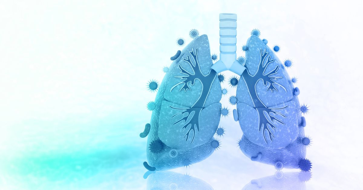
During the immediate post-operative period, ciliary dysfunction becomes pronounced as the delicate hair-like structures responsible for moving mucus become damaged or paralyzed. This mucociliary escalator malfunction allows infectious agents to establish biofilms within sinus cavities, leading to persistent inflammatory responses and recurrent bacterial overgrowth.
Primary Causes of Post-Septoplasty Sinus Infections
Surgical Trauma and Inflammatory Response
Post-operative inflammation represents the body’s natural healing response but simultaneously creates conditions favoring bacterial proliferation. Tissue edema compresses sinus drainage pathways, while inflammatory exudate provides nutrients for microbial growth.
The turbinate manipulation often performed alongside septoplasty further compromises nasal humidification and filtration functions. Reduced turbinate tissue decreases the nose’s ability to condition inspired air, allowing irritants and pathogens easier access to sinus cavities.
Altered Drainage Patterns
Septoplasty changes airflow dynamics within nasal passages, potentially redirecting air currents away from natural sinus drainage points. This aerodynamic alteration can create areas of stagnation where mucus accumulates, forming ideal breeding grounds for opportunistic pathogens.
Scar tissue formation during healing may permanently alter drainage angles, requiring months for the body to establish new, efficient clearance patterns. Some patients develop adhesions between nasal structures, further compromising drainage effectiveness.
Risk Factors for Increased Post-Surgical Infections
Patient-Specific Vulnerabilities
Certain individuals face elevated risks for post-septoplasty complications based on their medical history and anatomical variations. Understanding these predisposing factors helps predict and prevent infectious complications.
Table 1: Risk Factors for Post-Septoplasty Sinus Infections
| Risk Category | Specific Factors | Impact Level |
|---|---|---|
| Medical History | Chronic sinusitis, asthma, allergic rhinitis | High |
| Anatomical | Concha bullosa, paradoxical turbinates, narrow ostia | High |
| Immunological | Immunodeficiency, diabetes, chronic steroid use | High |
| Environmental | Smoking, occupational exposures, dry climate | Moderate |
| Surgical | Extensive resection, concurrent procedures, revision surgery | Moderate-High |
Pre-existing Sinus Disease
Patients with chronic rhinosinusitis before septoplasty face significantly higher infection rates post-operatively. The pre-existing inflammatory state creates a compromised healing environment where normal recovery processes become prolonged and complicated.
Allergic rhinitis patients experience heightened inflammatory responses that can persist weeks beyond typical healing timelines. The ongoing allergic cascade maintains mucosal edema and hypersecretion, perpetuating conditions favorable for bacterial overgrowth.
Timeline of Post-Septoplasty Complications
Immediate Post-Operative Period (0-2 weeks)
The first two weeks represent the highest risk period for infectious complications. Nasal packing removal, crusting formation, and peak inflammatory response create multiple opportunities for bacterial colonization.
During this phase, normal sinus drainage remains significantly impaired while surgical sites heal. Patients often experience thick, discolored nasal discharge that may indicate bacterial superinfection requiring antibiotic intervention.
Subacute Phase (2-8 weeks)
As initial healing progresses, some patients develop secondary infections characterized by persistent facial pressure, purulent drainage, and systemic symptoms. This period often reveals whether the patient will experience complicated versus uncomplicated recovery.
Crusting and synechiae formation during this phase can create permanent drainage impairments if not properly managed through regular follow-up care and appropriate interventions.
Long-term Recovery (2-6 months)
Most patients see gradual improvement in infection frequency as mucociliary function restores and drainage patterns normalize. However, some individuals require additional interventions to address persistent complications.
Prevention Strategies and Best Practices
Pre-operative Optimization
Successful infection prevention begins before surgery through comprehensive pre-operative evaluation and optimization. Addressing modifiable risk factors significantly reduces post-operative complication rates.
Pre-Surgical Checklist for Infection Prevention:
- Complete treatment of active sinus infections
- Optimize allergic rhinitis management
- Discontinue tobacco use minimum 4 weeks prior
- Address diabetes glucose control
- Review and adjust immunosuppressive medications
- Evaluate and treat nasal polyps or other concurrent pathology
Post-operative Care Protocols
Meticulous post-operative care represents the cornerstone of infection prevention. Adherence to prescribed irrigation regimens and follow-up schedules dramatically reduces complication rates.
Saline irrigation technique requires proper instruction and consistent execution. High-volume, low-pressure irrigation effectively removes debris and promotes healing without disrupting delicate surgical sites.
Table 2: Post-Operative Care Timeline and Interventions
| Time Period | Key Interventions | Infection Prevention Focus |
|---|---|---|
| 0-3 days | Antibiotic prophylaxis, gentle irrigation | Prevent primary bacterial seeding |
| 3-14 days | Regular debridement, intensive irrigation | Remove crusts and debris |
| 2-6 weeks | Steroid sprays, continued irrigation | Reduce inflammation, maintain patency |
| 6-12 weeks | Gradual activity resumption, monitoring | Detect and treat complications early |
Treatment Approaches for Post-Septoplasty Infections
Conservative Management
Most post-septoplasty sinus infections respond well to conservative treatment combining topical and systemic therapies. The key lies in early recognition and prompt intervention before complications develop.
Topical antibiotics delivered through irrigation solutions provide high local concentrations while minimizing systemic side effects. Mupirocin and gentamicin irrigations show particular efficacy against common post-surgical pathogens.
Medical Therapy Options
Systemic antibiotics remain the mainstay of treatment for established post-septoplasty sinus infections. Culture-guided therapy provides optimal results, though empirical coverage often proves necessary due to timing constraints.
Common Antibiotic Regimens for Post-Septoplasty Infections:
- First-line empirical therapy: Amoxicillin-clavulanate 875/125mg twice daily
- Penicillin-allergic patients: Doxycycline 100mg twice daily or azithromycin
- Severe infections: Levofloxacin 750mg daily or clindamycin plus third-generation cephalosporin
- Culture-guided therapy: Targeted based on sensitivity patterns
Adjunctive Treatments
Corticosteroids play a crucial role in managing the inflammatory component of post-septoplasty infections. Topical nasal steroids reduce mucosal edema and improve sinus drainage while systemic steroids may be necessary for severe cases.
Mucolytics and expectorants help thin secretions and promote clearance, particularly beneficial in patients with thick, tenacious mucus that impedes natural drainage mechanisms.
When to Seek Emergency Medical Care
Warning Signs and Red Flags – Get more sinus infections after septoplasty
Certain symptoms indicate serious complications requiring immediate medical evaluation. Delayed recognition of these warning signs can lead to devastating consequences including intracranial extension of infection.
Immediate Medical Attention Required:
- Severe headache with neck stiffness
- Visual changes or double vision
- High fever (>101.5°F) with systemic toxicity
- Facial swelling extending beyond surgical site
- Neurological symptoms or altered mental status
- Persistent bleeding or foul-smelling discharge
Serious Complications
Orbital cellulitis represents one of the most feared complications of post-septoplasty sinus infections. The thin lamina papyracea provides minimal barrier against infection spread from ethmoid sinuses to orbital contents.
Intracranial complications, though rare, can occur through direct extension or hematogenous spread. Meningitis, epidural abscess, and cavernous sinus thrombosis represent life-threatening sequelae requiring immediate hospitalization and aggressive treatment.
Long-term Outcomes and Prognosis
Recovery Expectations – Get more sinus infections after septoplasty
The vast majority of patients experiencing increased sinus infections after septoplasty see complete resolution within six months. Understanding normal healing timelines helps patients maintain realistic expectations during recovery.
Factors influencing long-term outcomes include pre-operative sinus health, adherence to post-operative care instructions, and prompt treatment of complications when they arise. Most patients ultimately achieve improved sinus function compared to their pre-operative state.
Success Rates and Patient Satisfaction
Studies demonstrate that 85-90% of septoplasty patients report improved nasal breathing and reduced sinus infection frequency by one year post-operatively. Even patients experiencing early complications typically achieve excellent long-term results with appropriate management.
Table 3: Long-term Outcomes After Septoplasty
| Outcome Measure | 6 Months | 1 Year | 2 Years |
|---|---|---|---|
| Improved nasal breathing | 75% | 85% | 88% |
| Reduced infection frequency | 70% | 82% | 85% |
| Patient satisfaction | 80% | 87% | 90% |
| Return to normal activities | 95% | 98% | 99% |
Expert Recommendations and Future Considerations
Optimizing Surgical Technique
Advances in endoscopic visualization and surgical instrumentation continue to reduce post-operative complication rates. Computer-assisted navigation systems help surgeons preserve critical anatomical structures while achieving optimal functional outcomes.
Minimally invasive techniques including balloon sinuplasty and tissue-sparing approaches show promise for reducing post-operative inflammation and subsequent infection risk. These evolving technologies may further improve patient outcomes in coming years.
Personalized Medicine Approaches
Future developments in genetic testing and biomarker analysis may allow surgeons to identify high-risk patients before surgery and customize treatment protocols accordingly. Personalized antibiotic prophylaxis based on individual microbiome analysis represents an exciting frontier in post-operative care.
Understanding individual variations in healing response and immune function will enable more targeted interventions, potentially eliminating post-septoplasty complications for many patients through precision medicine approaches.
Conclusion – Get more sinus infections after septoplasty
Increased sinus infections after septoplasty represent a common but typically temporary complication affecting roughly one in five patients during initial recovery. Understanding the underlying mechanisms, recognizing risk factors, and implementing appropriate prevention strategies significantly reduce both the incidence and severity of these infections.
Most patients experiencing post-septoplasty sinus infections achieve complete resolution within three to six months through conservative management and appropriate medical therapy. Early recognition of complications and prompt intervention remain crucial for optimal outcomes, while understanding when to seek emergency care prevents serious sequelae.
The long-term prognosis for septoplasty patients remains excellent, with the vast majority achieving improved nasal function and reduced infection rates compared to their pre-operative state. Continued advances in surgical technique and personalized medicine approaches promise even better outcomes for future patients undergoing this common procedure.
1. What are some common conditions that can be mistaken for a hernia?
Answer:
Several conditions can mimic the symptoms or appearance of a hernia, including:
Lipomas: Benign fatty lumps under the skin.
Lymphadenopathy: Swollen lymph nodes.
Hydrocele: Fluid-filled sac around the testicle.
Varicocele: Enlarged veins in the scrotum.
Muscle strain or tear: Can cause localized bulging or pain.
Abscess or cyst: Infected or fluid-filled lumps.
Tumors: Both benign and malignant growths.
2. How can a doctor differentiate between a hernia and other similar lumps?
Answer:
Doctors use a combination of physical examination and imaging studies to distinguish hernias from other conditions. Key methods include:
Physical exam: Checking for a bulge that enlarges with coughing or straining.
Ultrasound: Non-invasive imaging to visualize soft tissue structures.
CT scan or MRI: Detailed imaging if diagnosis is unclear.
Patient history: Symptoms such as pain, changes in size, or reducibility (whether the lump can be pushed back).
3. Can muscle strains or tears be confused with hernias?
Answer:
Yes, muscle strains or tears, especially in the abdominal or groin area, can cause localized swelling and pain that may resemble a hernia. However, muscle injuries usually do not produce a persistent bulge that changes with pressure or position, which is typical for hernias.
4. Are swollen lymph nodes ever mistaken for hernias?
Answer:
Swollen lymph nodes, particularly in the groin area, can sometimes be mistaken for hernias because they present as lumps. However, lymph nodes are usually firm, tender, and not reducible, and they do not change size with straining, unlike hernias.
5. What symptoms should prompt someone to see a doctor if they suspect a hernia?
Answer:
You should see a doctor if you notice:
A persistent lump or bulge in the abdomen, groin, or scrotum.
Pain or discomfort, especially when lifting, coughing, or bending.
A lump that increases in size or becomes tender.
Symptoms of bowel obstruction (nausea, vomiting, severe abdominal pain).
Redness or warmth over the lump, which could indicate incarceration or strangulation.
Early diagnosis helps prevent complications and ensures appropriate treatment.
Here are some common and notable conditions that can mimic a hernia:
Lipoma
Description: A benign tumor composed of fatty tissue. Lipomas are soft, usually movable, and generally painless. They can occur almost anywhere in the body, including the groin, abdominal wall, or spermatic cord, where they can be mistaken for hernias.
Why it’s mistaken: A lipoma in the inguinal canal or spermatic cord can present as a groin lump, similar to an inguinal hernia.
Evidence/Source:
Title: “Groin lumps: aetiology, diagnosis and management”
Authors: Jenkins, J. T., & O’Dwyer, P. J.
Source: British Journal of Hospital Medicine, 2008, 69(3), 141-145.
Excerpt/Relevance: This review article discusses various causes of groin lumps, including lipomas of the cord as a differential diagnosis for inguinal hernias.
Link: (Often behind a paywall, abstract usually available) https://www.magonlinelibrary.com/doi/abs/10.12968/hosp.2008.69.3.28819
Additional Evidence (StatPearls – good for summaries):
Title: Inguinal Hernia
Authors: Shamley, R. G.; Hira, K. G.; Ray, M. D. R.
Source: StatPearls Publishing; 2023 Jan.
Relevance: Lists lipoma in the differential diagnosis.
Link: https://www.ncbi.nlm.nih.gov/books/NBK459282/ (Search “lipoma” in the differential diagnosis section)
Lymphadenopathy (Swollen Lymph Nodes)
Description: Enlargement of lymph nodes, often due to infection, inflammation, or malignancy. Inguinal lymph nodes are located in the groin.
Why it’s mistaken: Swollen inguinal lymph nodes can present as firm lumps in the groin, potentially confused with an irreducible or incarcerated inguinal or femoral hernia.
Evidence/Source:
Title: “Evaluation of the adult with a groin lump”
Authors: Brooks, R. A., & Kurer, M. A. (Often covered in general surgical texts and review articles, this is a common clinical scenario rather than a specific study focusing only on this misdiagnosis). Many review articles on groin masses will list lymphadenopathy.
Source: This is a common topic in clinical guidelines and textbooks. For instance, the StatPearls article on Inguinal Hernia (cited above) also lists lymphadenopathy.
Specific Article:
Title: “A clinical review of an inguinal lymph node protocol”
Authors: Vezzosi, D., Bérouti, M., & Grunenwald, S., et al.
Source: BMC Cancer, 2017, 17, 676.
Relevance: While focused on a protocol, it inherently discusses the significance and diagnostic approach to inguinal lymphadenopathy, which is a key differential for groin masses.
Link: https://www.ncbi.nlm.nih.gov/pmc/articles/PMC5627434/
Hydrocele / Spermatocele / Epididymal Cyst
Description:
Hydrocele: A collection of fluid around a testicle.
Spermatocele: A painless, fluid-filled cyst in the epididymis, usually containing nonviable sperm.
Epididymal cyst: Similar to a spermatocele but contains clear fluid instead of sperm.
Why it’s mistaken: These present as scrotal swellings that can sometimes extend towards the inguinal region or be confused with an inguinoscrotal hernia. A hydrocele of the cord can present as a lump in the inguinal canal.
Evidence/Source:
Title: “Scrotal Swellings”
Authors: Leslie, S.W., Sajjad, H., Siref, L.E.
Source: StatPearls Publishing; 2023 Jan.
Relevance: This article provides a comprehensive overview of scrotal swellings and their differential diagnoses, which often include hernias.
Link: https://www.ncbi.nlm.nih.gov/books/NBK441973/
Varicocele
Description: Enlargement of the veins within the scrotum (pampiniform plexus). Often described as feeling like a “bag of worms.”
Why it’s mistaken: While typically distinct on palpation, a large varicocele might cause a sense of fullness or a visible bulge in the scrotal/inguinal area, which could be confused with a hernia, especially if pain is present.
Evidence/Source:
The StatPearls article “Inguinal Hernia” (Shamley et al., cited above) lists varicocele in its differential diagnosis.
Title: “Varicocele”
Authors: Leslie, S.W., Sajjad, H., & Cathey, C.
Source: StatPearls Publishing; 2023 Jan.
Relevance: Details varicocele, and its presentation can sometimes overlap with hernia concerns for patients.
Undescended Testis (Cryptorchidism)
Description: A testis that has not moved into its proper position in the scrotum before birth. It may be located in the abdomen or, more commonly, in the inguinal canal.
Why it’s mistaken: An undescended testis palpable in the inguinal canal presents as a lump and can be mistaken for an inguinal hernia, especially in infants and children. Often, a hernia coexists.
Evidence/Source:
Title: “Undescended Testis (Cryptorchidism)”
Authors: Leslie, S.W., Sajjad, H., & Kasi, A.
Source: StatPearls Publishing; 2023 Jan.
Relevance: Discusses the presentation of undescended testes, including palpation in the inguinal canal.
Kiểm Duyệt Nội Dung
More than 10 years of marketing communications experience in the medical and health field.
Successfully deployed marketing communication activities, content development and social networking channels for hospital partners, clinics, doctors and medical professionals across the country.
More than 6 years of experience in organizing and producing leading prestigious medical programs in Vietnam, in collaboration with Ho Chi Minh City Television (HTV). Typical programs include Nhật Ký Blouse Trắng, Bác Sĩ Nói Gì, Alo Bác Sĩ Nghe, Nhật Ký Hạnh Phúc, Vui Khỏe Cùng Con, Bác Sỹ Mẹ, v.v.
Comprehensive cooperation with hundreds of hospitals and clinics, thousands of doctors and medical experts to join hands in building a medical content and service platform on the Doctor Network application.


