Sinus infections affect millions of people annually, typically causing manageable symptoms like nasal congestion and facial pressure. However, in extremely rare cases—occurring in less than 1% of sinusitis patients—these common infections can escalate into life-threatening medical emergencies when bacteria breach the natural barriers between your sinuses and brain. This neurological complication represents one of medicine’s most critical urgent care scenarios, requiring immediate hospital intervention to prevent permanent brain damage, stroke, or death.
How to Tell If a Sinus Infection Has Spread to Brain – This comprehensive guide will examine the anatomical pathways enabling bacterial spread, identify specific neurological warning signs that distinguish dangerous complications from routine sinusitis symptoms, explore diagnostic procedures used by emergency physicians, and outline treatment protocols for these severe infections. Understanding these critical distinctions could save your life or that of a loved one when minutes matter most.
Understanding Sinus Infections: Essential Background

Sinusitis occurs when the hollow air-filled cavities surrounding your nasal passages become inflamed due to viral, bacterial, or fungal pathogens. These paranasal sinuses—including the frontal, ethmoid, sphenoid, and maxillary chambers—normally drain mucus through narrow openings into the nasal cavity. When inflammation blocks these drainage pathways, trapped secretions create ideal breeding conditions for harmful microorganisms.
Typical acute sinusitis manifests through recognizable symptoms: nasal obstruction, purulent discharge, facial pain or pressure, reduced sense of smell, cough, and general malaise. Most cases resolve within 7-10 days with appropriate treatment, including decongestants, saline irrigation, and sometimes antibiotics for bacterial infections.
The key distinction lies in symptom severity and neurological involvement. Standard sinus infections remain localized within the sinus cavities, while dangerous complications occur when pathogens cross anatomical boundaries to invade surrounding structures.
Why and How Sinus Infections Spread: Understanding the Mechanism
The proximity between your sinuses and brain creates potential pathways for bacterial invasion during severe infections. These anatomical connections explain why certain sinus infections can become neurosurgical emergencies requiring immediate intervention.
Direct Extension Through Bone
Aggressive bacterial strains can erode through the thin bony walls separating sinuses from intracranial structures. The ethmoid and sphenoid sinuses pose particular risks due to their close proximity to the brain’s frontal lobes and skull base. Osteomyelitis—bone infection—can develop when bacteria destroy these protective barriers.
Vascular Spread
The cavernous sinus, a major venous structure at the skull base, receives drainage from facial veins and connects directly to brain circulation. Bacterial emboli can travel through these vessels, causing cavernous sinus thrombosis—a potentially fatal condition involving blood clot formation in this critical area.
Neural Pathway Invasion
Cranial nerves passing through sinus regions provide another route for bacterial spread. The olfactory nerve, which enables smell sensation, extends directly from the nasal cavity into the brain, creating a potential pathway for pathogen migration.
This spread becomes dangerous because it introduces infection into sterile areas where the body’s immune defenses are limited. Brain tissue lacks the robust inflammatory response found elsewhere, making infections particularly destructive once established.
Key Warning Signs and Symptoms of Brain Involvement
Recognizing neurological symptoms that signal dangerous complications requires understanding how brain infection differs from routine sinusitis. These warning signs indicate that bacteria have breached normal anatomical boundaries and begun affecting central nervous system function.
Critical Neurological Symptoms
Severe, Unrelenting Headache Brain involvement typically produces excruciating headaches that differ markedly from typical sinus pressure. These headaches often worsen progressively, resist standard pain medications, and may be accompanied by photophobia (light sensitivity) or phonophobia (sound sensitivity). The pain results from increased intracranial pressure or direct inflammatory irritation of brain tissue.
High-Grade Fever with Rigors Temperature elevation above 101.3°F (38.5°C) accompanied by severe shaking chills suggests systemic bacterial spread beyond localized sinus infection. This fever pattern indicates that pathogens have entered the bloodstream or established infection in deeper tissues.
Visual Disturbances Double vision (diplopia), blurred vision, or visual field defects can result from cranial nerve compression or cavernous sinus involvement. The optic nerve and muscles controlling eye movement may become affected when infection spreads to surrounding structures.
Periorbital Swelling and Erythema Significant eyelid swelling, particularly when accompanied by redness and warmth, may indicate orbital cellulitis—a serious complication often preceding intracranial spread. This occurs most commonly with ethmoid sinusitis due to the shared blood supply between the orbit and these sinus cavities.
Table 1: Comparison of Standard Sinusitis vs. Brain Involvement Symptoms
| Symptom Category | Standard Sinusitis | Brain Involvement |
|---|---|---|
| Headache | Mild to moderate facial pressure | Severe, progressive, unrelenting |
| Fever | Low-grade or absent | High fever >101.3°F (38.5°C) |
| Mental Status | Normal alertness | Confusion, altered consciousness |
| Vision | Unaffected | Double vision, visual field defects |
| Neurological | No deficits | Weakness, numbness, speech changes |
| Neck | Normal flexibility | Stiffness, pain with movement |
Neck Stiffness (Nuchal Rigidity) Inability to flex the neck forward without severe pain represents a classic sign of meningitis—infection of the protective membranes surrounding the brain and spinal cord. This symptom results from inflammatory irritation of the meninges and requires immediate medical evaluation.
Altered Mental Status Confusion, disorientation, memory problems, or changes in personality indicate that infection has begun affecting brain function directly. These cognitive changes may progress rapidly from subtle confusion to complete loss of consciousness.
Neurological Deficits Weakness, numbness, or paralysis affecting one side of the body suggests brain abscess formation or stroke secondary to infection. Speech difficulties, including slurred words or inability to find appropriate language, indicate involvement of specific brain regions controlling communication.
Seizure Activity New-onset seizures in the context of sinus infection represent a neurological emergency. Seizures occur when bacterial toxins or inflammatory mediators irritate brain tissue, causing abnormal electrical activity.
These symptoms represent medical emergencies requiring immediate hospital evaluation. The presence of any neurological symptom alongside sinus infection warrants urgent medical attention, as delayed treatment significantly increases mortality and morbidity risks.
Specific Life-Threatening Complications
When sinus infections spread beyond their normal boundaries, several distinct but equally dangerous conditions can develop. Each represents a neurosurgical emergency requiring specialized treatment protocols and intensive monitoring.
Bacterial Meningitis
Meningitis involves infection of the cerebrospinal fluid and protective membranes (meninges) surrounding the brain and spinal cord. Bacteria reach these sterile spaces through direct extension from infected sinuses or hematogenous spread through the bloodstream.
Clinical presentation includes the classic triad of fever, headache, and neck stiffness, often accompanied by photophobia and altered mental status. Bacterial meningitis can progress rapidly from initial symptoms to coma and death within hours, making immediate antibiotic treatment crucial for survival.
Brain Abscess
A brain abscess represents a localized collection of pus within brain tissue, typically forming when bacteria establish infection in a specific brain region. These lesions can expand rapidly, causing increased intracranial pressure and mass effect on surrounding structures.
Symptoms depend on abscess location but commonly include progressive headache, focal neurological deficits, and seizures. Frontal lobe abscesses may cause personality changes and motor weakness, while temporal lobe involvement can affect speech and memory function.
Cavernous Sinus Thrombosis
This rare but life-threatening condition occurs when blood clots form within the cavernous sinus—a major venous structure at the skull base. Bacteria spreading from sphenoid or ethmoid sinuses can cause inflammation leading to thrombosis.
Characteristic features include severe headache, high fever, and cranial nerve palsies affecting eye movement and facial sensation. Patients often develop proptosis (eye protrusion) and chemosis (conjunctival swelling) due to impaired venous drainage.
Subdural Empyema
Subdural empyema involves pus collection between the brain’s protective layers—specifically between the dura mater and arachnoid membrane. This condition typically results from direct extension of frontal or ethmoid sinusitis through the skull base.
The clinical course often includes rapid neurological deterioration with seizures, focal deficits, and decreased consciousness. Emergency surgical drainage combined with prolonged antibiotic therapy is usually required for successful treatment.
Table 2: Intracranial Complications – Key Features and Mortality Rates
| Complication | Primary Location | Mortality Rate | Key Clinical Features |
|---|---|---|---|
| Bacterial Meningitis | Cerebrospinal fluid | 10-15% | Fever, headache, neck stiffness |
| Brain Abscess | Brain parenchyma | 5-10% | Focal deficits, seizures, headache |
| Cavernous Sinus Thrombosis | Venous sinuses | 15-20% | Eye movement problems, facial swelling |
| Subdural Empyema | Subdural space | 10-20% | Rapid deterioration, seizures |
Orbital Cellulitis
While not technically intracranial, orbital cellulitis represents a serious complication that can precede or accompany brain involvement. This infection affects the soft tissues within the eye socket, typically spreading from ethmoid or frontal sinuses.
Signs include marked eyelid swelling, eye pain, restricted eye movement, and potential vision loss. Orbital cellulitis can progress to cavernous sinus thrombosis or intracranial extension if not treated promptly with intravenous antibiotics.
Risk Factors: Who Is Most Vulnerable?
Understanding individual risk factors helps identify patients most likely to develop serious complications from sinus infections. While these severe outcomes remain rare in the general population, certain conditions significantly increase vulnerability to bacterial spread.
Immunocompromised States
Patients with weakened immune systems face dramatically increased risks for complicated sinusitis. Diabetes mellitus creates an environment favorable to bacterial growth while impairing the body’s ability to fight infection effectively. HIV/AIDS patients, organ transplant recipients, and individuals receiving chemotherapy or immunosuppressive medications all demonstrate higher complication rates.
Chronic corticosteroid use, whether for asthma, arthritis, or other inflammatory conditions, suppresses local and systemic immune responses. These patients may develop fulminant infections with minimal initial symptoms, making early recognition particularly challenging.
Anatomical Variations and Prior Surgery
Certain anatomical variations can predispose individuals to both recurrent sinusitis and increased complication risks. Deviated nasal septum, concha bullosa, or narrow sinus ostia can impair normal drainage, creating conditions favorable for bacterial overgrowth.
Previous sinus surgery, while often helpful for chronic sinusitis, can occasionally create scar tissue or anatomical changes that alter normal drainage patterns. Endoscopic procedures may rarely cause inadvertent communication between sinuses and intracranial spaces.
Chronic Medical Conditions
Chronic rhinosinusitis patients experience repeated episodes of inflammation and infection, potentially leading to progressive tissue damage and increased susceptibility to complications. The presence of nasal polyps, cystic fibrosis, or primary ciliary dyskinesia further impairs normal sinus function.
Age-Related Factors
Very young children and elderly adults demonstrate increased vulnerability to serious infections. Pediatric patients have incompletely developed immune systems and thinner skull bones, while elderly individuals often have multiple medical comorbidities affecting immune function.
Environmental and Behavioral Factors
- Delayed medical treatment for initial sinus symptoms
- Inadequate antibiotic treatment or poor medication compliance
- Recent dental infections or procedures affecting maxillary sinuses
- Recreational drug use, particularly cocaine, which can cause severe nasal and sinus damage
- Swimming or diving activities that may force contaminated water into sinus cavities
Diagnostic Evaluation: How Medical Professionals Identify Spread
When patients present with concerning symptoms suggesting intracranial complications, emergency physicians and specialists employ a systematic diagnostic approach combining clinical assessment with advanced imaging and laboratory studies.
Emergency Department Assessment
The initial evaluation begins with a focused history and physical examination. Physicians assess the timeline of symptom development, previous treatments attempted, and any underlying medical conditions. The neurological examination includes assessment of mental status, cranial nerve function, motor strength, reflexes, and signs of meningeal irritation.
Vital signs provide crucial information about infection severity. High fever, elevated heart rate, and changes in blood pressure may indicate systemic infection or increased intracranial pressure.
Advanced Imaging Studies
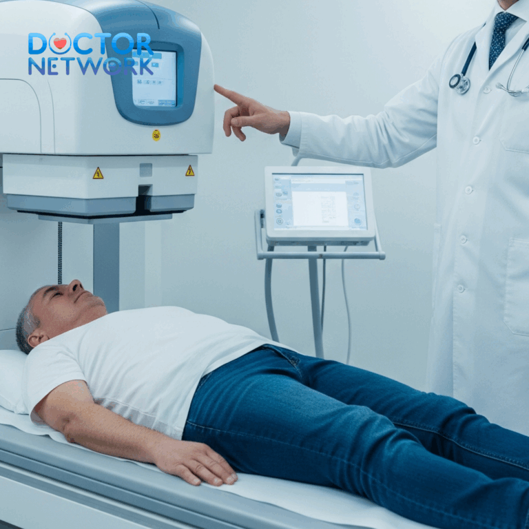
Computed Tomography (CT) Scanning CT imaging of the sinuses and brain represents the initial imaging study of choice in emergency settings. Modern CT scanners can rapidly identify sinus opacification, fluid levels, and bony destruction while simultaneously evaluating for intracranial complications.
CT findings suggesting bacterial spread include:
- Sinus wall erosion or destruction
- Orbital soft tissue involvement
- Intracranial fluid collections
- Brain tissue enhancement after contrast administration
- Signs of increased intracranial pressure
Magnetic Resonance Imaging (MRI) MRI provides superior soft tissue contrast and better visualization of intracranial complications. This study is particularly valuable for detecting early brain abscess formation, evaluating the extent of meningeal enhancement, and assessing for cavernous sinus involvement.
MRI sequences specifically designed for infection evaluation can identify inflammatory changes before they become apparent on CT imaging.
Laboratory Studies
Blood Cultures and Inflammatory Markers Blood cultures help identify causative organisms and guide antibiotic selection. Complete blood count typically reveals elevated white blood cell count with increased neutrophil percentage (left shift). Inflammatory markers including C-reactive protein and erythrocyte sedimentation rate are usually markedly elevated.
Lumbar Puncture (Spinal Tap) When meningitis is suspected, lumbar puncture provides definitive diagnosis through cerebrospinal fluid analysis. This procedure must be performed carefully after ensuring no evidence of increased intracranial pressure that might make the procedure dangerous.
Cerebrospinal fluid findings in bacterial meningitis include:
- Elevated protein levels
- Decreased glucose concentration
- Increased white blood cell count with neutrophil predominance
- Positive bacterial cultures or antigen testing
Microbiological Studies
Sinus Aspiration and Culture Direct sampling of sinus contents through endoscopic aspiration provides the most accurate identification of causative organisms. This procedure helps guide targeted antibiotic therapy and identifies antibiotic-resistant bacteria.
Molecular Diagnostic Testing Advanced techniques including polymerase chain reaction (PCR) testing can rapidly identify bacterial DNA even when traditional cultures are negative. These methods are particularly valuable for patients who have received partial antibiotic treatment.
Treatment Approaches: Emergency Management and Long-Term Care
Managing intracranial complications from sinus infections requires immediate, aggressive treatment combining high-dose intravenous antibiotics with surgical intervention when necessary. Treatment decisions must be made rapidly to prevent irreversible neurological damage.
Emergency Antibiotic Therapy
Initial Empirical Treatment Before culture results become available, broad-spectrum antibiotics targeting the most likely pathogens must be initiated immediately. The choice of antibiotics considers common sinusitis bacteria while ensuring adequate brain penetration.
Typical empirical regimens include:
- Vancomycin plus Ceftriaxone or Cefepime for broad coverage
- Metronidazole added for anaerobic bacteria coverage
- Ampicillin may be included for Listeria coverage in certain populations
These medications are administered intravenously at maximum recommended doses to achieve therapeutic levels in infected brain tissue.
Targeted Therapy Once culture results and antibiotic sensitivities become available, treatment is narrowed to the most effective agents for identified organisms. Treatment duration typically ranges from 4-8 weeks depending on the specific complication and patient response.
Surgical Interventions
Neurosurgical Procedures Brain abscesses often require surgical drainage through stereotactic aspiration or open craniotomy. The choice of procedure depends on abscess size, location, and patient neurological status. Smaller abscesses may respond to antibiotic therapy alone, while larger lesions typically require surgical intervention.
Subdural empyema almost always necessitates emergency surgical drainage to prevent rapid neurological deterioration. Delay in surgical treatment significantly increases mortality and morbidity risks.
Sinus Surgery Functional endoscopic sinus surgery (FESS) may be necessary to establish adequate drainage and remove infected tissue from the primary sinus infection. This procedure helps prevent recurrent complications while allowing better antibiotic penetration.
Emergency sinus surgery is sometimes required to drain purulent material and reduce bacterial load before addressing intracranial complications.
Supportive Care Measures
Intracranial Pressure Management Patients with brain abscesses or cerebral edema may require medications to reduce intracranial pressure. Osmotic agents like mannitol or hypertonic saline can provide temporary pressure relief while definitive treatment takes effect.
Seizure Control Anti-epileptic medications are often necessary for patients who develop seizures. Prophylactic anticonvulsants may be considered for high-risk patients even without seizure activity.
Intensive Care Monitoring Most patients with intracranial complications require intensive care unit admission for continuous neurological monitoring and support of vital functions.
Table 3: Treatment Duration and Monitoring Requirements
| Complication | Antibiotic Duration | Surgical Requirements | Follow-up Imaging |
|---|---|---|---|
| Meningitis | 2-3 weeks IV | Rarely needed | CT at completion |
| Brain Abscess | 6-8 weeks | Often required | MRI weekly initially |
| Cavernous Sinus Thrombosis | 3-4 weeks | Sometimes needed | MRI/MRV follow-up |
| Subdural Empyema | 4-6 weeks | Almost always | CT/MRI serial imaging |
Prevention Strategies: Reducing Complication Risks
While severe complications remain rare, implementing appropriate prevention strategies can significantly reduce the likelihood of dangerous bacterial spread from routine sinus infections.
Early Medical Intervention
Recognizing Concerning Symptoms Seeking medical attention promptly when sinus infection symptoms persist beyond 7-10 days or worsen after initial improvement helps identify bacterial infections requiring antibiotic treatment. Signs warranting medical evaluation include:
- Purulent nasal discharge lasting more than one week
- Facial pain or pressure that worsens rather than improves
- Fever developing after several days of illness
- Symptoms that improve then suddenly worsen
Appropriate Antibiotic Use When bacterial sinusitis is diagnosed, completing the full prescribed antibiotic course prevents treatment failure and reduces bacterial resistance development. Patients should not discontinue antibiotics early even if symptoms improve.
Managing Underlying Risk Factors
Diabetes Control Maintaining optimal blood glucose levels helps preserve immune function and reduces infection risks. Diabetic patients should monitor blood sugars more frequently during any infection and seek medical attention for persistent symptoms.
Immunocompromised Patient Care Individuals with weakened immune systems should maintain close communication with their healthcare providers and seek evaluation promptly for any signs of infection. Prophylactic measures may be appropriate for high-risk patients.
Proper Sinus Hygiene
Nasal Irrigation Regular saline irrigation helps maintain healthy sinus drainage and may reduce infection frequency. Using sterile or properly boiled water prevents introducing harmful organisms into sinus cavities.
Environmental Modifications
- Using humidifiers to maintain adequate humidity levels
- Avoiding known allergens that trigger sinus inflammation
- Practicing good hand hygiene to prevent viral infections
- Avoiding tobacco smoke and other respiratory irritants
Dental Health Maintenance
Since maxillary sinus infections can originate from dental sources, maintaining good oral hygiene and addressing dental problems promptly helps prevent sinus complications.
Conclusion – How to Tell If a Sinus Infection Has Spread to Brain

Sinus infections represent common medical conditions affecting millions annually, with most cases resolving without complications through appropriate treatment. However, the rare progression to intracranial involvement creates life-threatening emergencies requiring immediate recognition and aggressive intervention.
The key to preventing tragedy lies in understanding the critical warning signs that distinguish dangerous complications from routine sinusitis symptoms. Severe headache, high fever, visual changes, neck stiffness, altered mental status, and neurological deficits all signal potential brain involvement requiring emergency medical evaluation.
Healthcare professionals employ sophisticated diagnostic tools including advanced imaging and laboratory studies to identify bacterial spread rapidly. Treatment combines high-dose intravenous antibiotics with surgical intervention when necessary, often requiring intensive care support and prolonged therapy.
Prevention strategies focusing on early medical intervention, appropriate antibiotic use, and management of underlying risk factors can significantly reduce complication risks. Patients with compromised immune systems, diabetes, or chronic sinus conditions should maintain particularly close communication with their healthcare providers.
Critical Reminder: This information serves educational purposes and cannot substitute for professional medical evaluation. Any individual experiencing severe symptoms alongside sinus infection should seek immediate emergency medical attention. When neurological symptoms develop, minutes can determine the difference between complete recovery and permanent disability or death.
The rarity of these complications should not diminish awareness of their potential severity. Understanding these warning signs empowers patients and families to recognize medical emergencies and seek life-saving treatment when every moment counts.
5 frequently asked questions
1. Can a sinus infection spread to the brain?
Yes, although it is rare, a sinus infection can spread to the brain because the sinuses are located close to the brain and separated only by thin bones. When the infection breaches these barriers, it can cause serious complications such as meningitis, encephalitis, or brain abscesses.
2. What are the warning signs that a sinus infection has spread to the brain?
Signs include severe headache that is worse than typical sinus pain, high fever, neck stiffness, nausea or vomiting, sensitivity to bright light, vision changes (such as double or blurred vision), confusion or changes in mental status, seizures, muscle weakness or partial paralysis, personality changes, and loss of consciousness.
3. How urgent is it to seek medical help if these symptoms appear?
It is a medical emergency. Anyone experiencing these symptoms alongside a sinus infection should seek immediate medical attention by going to the emergency room or calling emergency services, as brain involvement can be life-threatening without prompt treatment.
4. How do doctors diagnose if a sinus infection has spread to the brain?
Doctors use a combination of physical and neurological examinations to assess vision, hearing, speech, coordination, and mental status. Imaging tests like CT scans or MRI are performed to detect inflammation, abscesses, or lesions in the brain. A lumbar puncture (spinal tap) may be done to analyze cerebrospinal fluid for infection.
5. What treatments are available if a sinus infection has spread to the brain?
Treatment depends on the specific complication but generally includes intravenous antibiotics or antifungal medications to fight infection, corticosteroids to reduce brain inflammation, and sometimes surgical drainage of the sinuses or brain abscesses. Anticonvulsants may be used to control seizures. Early treatment improves outcomes and reduces the risk of permanent damage.
References
General Reviews on Intracranial Complications of Sinusitis:
Title: “Intracranial Complications of Sinusitis”
Authors: Clayman, G. L., Adams, G. L., Paugh, D. R., & Koopmann Jr, C. F.
Source: Laryngoscope, 1991.
Key Points: An older but foundational review discussing the routes of spread (direct extension, retrograde thrombophlebitis), common pathogens, and types of complications (meningitis, various abscesses). It emphasizes the importance of early diagnosis and aggressive treatment. It highlights symptoms like persistent headache, fever, vomiting, lethargy, and focal neurological signs.
Link (PubMed): https://pubmed.ncbi.nlm.nih.gov/2046601/
DOI: 10.1288/00005537-199106000-00003
Title: “Intracranial complications of rhinosinusitis: a review”
Authors: Maniglia, A. J., Goodwin, W. J., Arnold, J. E., & Ganz, E.
Source: Laryngoscope, 1989.
Key Points: Another classic review detailing the pathophysiology, diagnosis, and management of these severe complications, emphasizing the warning signs.
Link (PubMed): https://pubmed.ncbi.nlm.nih.gov/2669864/
DOI: 10.1288/00005537-198907000-00001
Studies Focusing on Specific Complications and Their Presentation:
Brain Abscess:
Title: “Brain abscess secondary to sinusitis”
Authors: Roche, M., Humphreys, H., Smyth, E., Phillips, J., & Cunney, R.
Source: Journal of Antimicrobial Chemotherapy, 2003 (letter/brief report but part of a larger body of evidence).
Key Points: While a brief report, it underscores sinusitis as a cause of brain abscess. Larger case series and reviews consistently list headache, fever, and focal neurological deficits as key symptoms.
Link (Full Text via Oxford Academic): https://academic.oup.com/jac/article/51/1/235/716174
DOI: 10.1093/jac/dkg030
Subdural Empyema:
Title: “Subdural empyema of sinogenic origin: a case series and review of the literature”
Authors: Germiller, J. A., Monin, D. L., Sparano, A. M., & Tom, L. W. C.
Source: International Journal of Pediatric Otorhinolaryngology, 2006.
Key Points: This study reviews cases of subdural empyema originating from sinusitis, particularly in children and adolescents. Symptoms highlighted include headache, fever, altered mental status, seizures, and focal neurological deficits.
Link (PubMed): https://pubmed.ncbi.nlm.nih.gov/16458341/
DOI: 10.1016/j.ijporl.2005.11.006
Cavernous Sinus Thrombosis:
Title: “Cavernous sinus thrombosis: A review.”
Authors: Cannon, M. L., Antonio-Santos, A. A., Hart, D., Howard, J., & Winter, T.
Source: Clinical Ophthalmology, 2009 (This is a systematic review, but the actual published version is often cited as by various authors from Wills Eye Institute). A more general, accessible one:
A more accessible review:
Title: “Cavernous Sinus Thrombosis.”
Authors: Plewa, M.C., Tadi, P., & Gupta, M.
Source: StatPearls [Internet]. Treasure Island (FL): StatPearls Publishing; 2024 Jan-. (Continuously updated peer-reviewed book).
Key Points: Describes CST as a rare but life-threatening condition. Symptoms include severe headache, periorbital edema (swelling around the eye), proptosis (bulging eye), ophthalmoplegia (inability to move the eye), vision loss, fever, and sepsis. Sinusitis is a common cause.
Link (NCBI Bookshelf – Full Text): https://www.ncbi.nlm.nih.gov/books/NBK448164/
Modern Case Series and Diagnostic Approach:
Title: “Intracranial Complications of Acute Rhinosinusitis: A Retrospective Case Series”
Authors: Bayonne, E., Kania, R., Tran, P., Huy, B., & Herman, P.
Source: The Laryngoscope, 2009.
Key Points: Reviews 20 cases, highlighting common symptoms like headache (100%), fever (70%), and neurological signs (60%). Emphasizes the need for CT and MRI for diagnosis, and combined medical and surgical treatment.
Link (PubMed): https://pubmed.ncbi.nlm.nih.gov/19260098/
DOI: 10.1002/lary.20140
Kiểm Duyệt Nội Dung
More than 10 years of marketing communications experience in the medical and health field.
Successfully deployed marketing communication activities, content development and social networking channels for hospital partners, clinics, doctors and medical professionals across the country.
More than 6 years of experience in organizing and producing leading prestigious medical programs in Vietnam, in collaboration with Ho Chi Minh City Television (HTV). Typical programs include Nhật Ký Blouse Trắng, Bác Sĩ Nói Gì, Alo Bác Sĩ Nghe, Nhật Ký Hạnh Phúc, Vui Khỏe Cùng Con, Bác Sỹ Mẹ, v.v.
Comprehensive cooperation with hundreds of hospitals and clinics, thousands of doctors and medical experts to join hands in building a medical content and service platform on the Doctor Network application.


