Sebaceous cyst in the scrotum – Scrotal sebaceous cysts are benign, encapsulated nodules that develop on the scrotal skin when sebaceous glands become blocked, leading to the accumulation of keratin material and sebum within a subepidermal sac. These firm, dome-shaped protrusions represent one of the most common dermatological conditions affecting the male genital area, occurring when the microscopic oil-producing glands in the scrotal tissue fail to drain properly. While typically harmless and painless, these retention cysts can cause significant psychological distress, cosmetic concerns, and occasionally develop complications requiring medical intervention.
This comprehensive guide explores the anatomical basis of scrotal sebaceous cyst formation, diagnostic approaches, treatment modalities, and long-term management strategies. We’ll examine the distinguishing characteristics that differentiate these benign growths from other scrotal masses, discuss both conservative and surgical treatment options, and address the psychological impact these conditions can have on patients. Additionally, we’ll cover prevention strategies, potential complications including infection risks, and the excellent prognosis associated with appropriate medical care.
Understanding the Scrotum and Sebaceous Glands
The scrotum functions as a protective sac of skin positioned beneath the penis, housing and safeguarding the testicles while maintaining the optimal temperature necessary for spermatogenesis. This anatomical structure also contains the epididymis, an elongated tubular structure responsible for sperm storage and maturation. The scrotal skin contains numerous sebaceous glands, microscopic structures that produce sebum—a thick, oily substance essential for skin lubrication and hair maintenance.
Sebaceous glands discharge their oily secretions through specialized ducts that connect to hair follicles throughout the scrotal region. When these ducts become obstructed due to cellular debris, trauma, or developmental abnormalities, sebum accumulates within the gland, creating a retention cyst. This blockage mechanism explains why scrotal sebaceous cysts often develop in areas with dense hair follicle concentration.
The formation process begins when keratinocytes and sebaceous material cannot properly exit through the follicular opening. Over time, this trapped material expands the gland structure, creating the characteristic firm, encapsulated nodule that defines a sebaceous cyst. Understanding this pathophysiology helps explain why complete surgical removal of the cyst wall prevents recurrence, while simple drainage often leads to reformation.

Causes and Risk Factors
Blocked sebaceous gland ducts represent the primary etiology behind scrotal sebaceous cyst development. Several factors contribute to this obstruction, including developmental abnormalities in sebaceous duct formation and traumatic injury that forces surface epithelial cells deeper into the dermal layers. Genetic predisposition plays a significant role, particularly in individuals with Gardner syndrome, an inherited condition associated with multiple cyst formation throughout the body.
Certain behavioral factors can increase the likelihood and severity of cyst development. Attempting home removal through cutting or squeezing often exacerbates the condition, enlarging existing cysts while increasing infection risk and causing permanent scarring. Poor wound healing from these DIY attempts can create additional blockage points, leading to multiple cyst formation.
Hair removal practices, particularly aggressive shaving and waxing of the scrotal area, may contribute to cyst development by creating ingrown hairs and follicular damage. These mechanical insults can disrupt normal sebaceous gland drainage, predisposing individuals to retention cyst formation. However, complete prevention remains challenging, as some cases result from congenital duct abnormalities or spontaneous blockage despite optimal hygiene practices.
Appearance and Characteristics
Scrotal sebaceous cysts present as whitish, yellowish, or skin-colored lumps filled with clear oily liquid or putty-like keratinous material. These dome-shaped protrusions vary considerably in size, ranging from tiny 1-2 millimeter nodules to larger 1-2 centimeter masses, with some patients presenting pea-sized lesions alongside larger growths simultaneously.
The distribution pattern often involves multiple cysts scattered across both sides of the scrotal midline, though solitary lesions also occur. In severe cases, numerous cysts may cover the entire scrotal surface, creating an appearance resembling cauliflower or grape clusters. This extensive involvement can significantly impact the scrotal contour and cause considerable cosmetic distress.
| Characteristic | Typical Presentation | Variations |
|---|---|---|
| Color | Whitish to yellowish | May match surrounding skin tone |
| Size | 1-2mm to 2cm diameter | Can vary within same patient |
| Number | Often multiple | Single cysts possible |
| Distribution | Bilateral scrotal involvement | May concentrate on one side |
| Texture | Firm, dome-shaped | Consistency varies with contents |
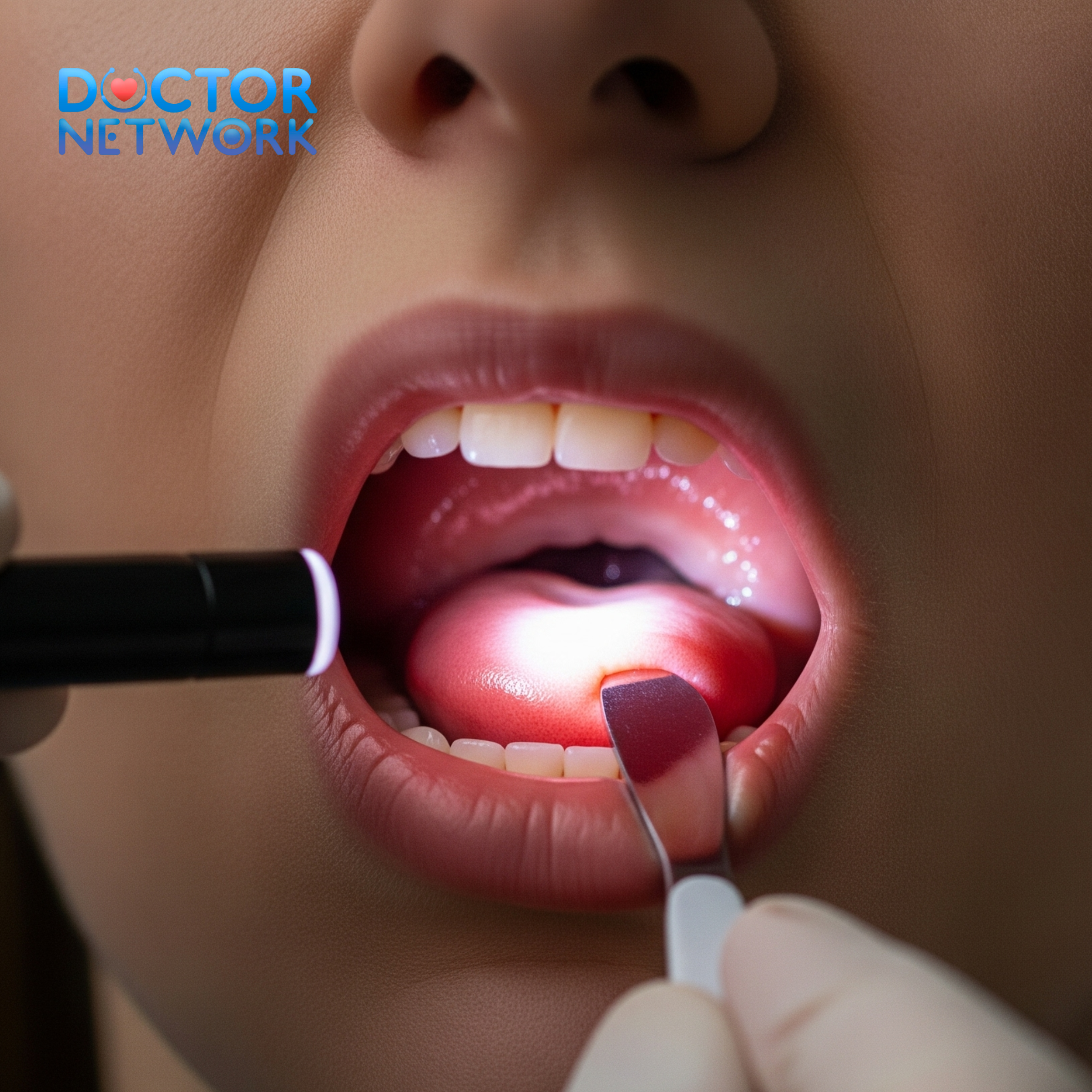
Most cysts maintain consistent size over time, though some may gradually enlarge. The cyst contents typically include accumulated sebum, keratin debris, and occasionally calcified material in long-standing lesions. Transillumination with a light source may reveal the cystic nature of smaller lesions, helping distinguish them from solid masses.
Symptoms and When to Seek Medical Attention
The majority of scrotal sebaceous cysts remain asymptomatic, presenting as painless nodules discovered during routine self-examination or partner observation. However, patients may experience various symptoms depending on cyst size, location, and individual sensitivity factors.
Common symptoms include a sensation of heaviness in the scrotal region, particularly when multiple large cysts are present. Some individuals report dull, aching discomfort rather than sharp pain, along with mild swelling around affected areas. The presence of visible lumps often causes significant embarrassment and anxiety, impacting self-confidence and intimate relationships.
Signs requiring immediate medical evaluation include:
- Sudden onset of severe scrotal or testicular pain
- Rapid cyst enlargement or firmness changes
- Redness, warmth, or tenderness around cysts
- Purulent discharge with foul odor
- Fever accompanying scrotal symptoms
- Any new or unexplained scrotal masses
Infected cysts become painful and may spontaneously rupture, discharging malodorous pus. This complication requires prompt antibiotic treatment to prevent spreading infection. Additionally, any sudden, severe testicular pain warrants emergency evaluation to exclude testicular torsion, a urological emergency requiring immediate surgical intervention.
Patients should seek professional assessment for recurrent cysts, particularly those showing changes in size, appearance, or associated symptoms. Early medical consultation helps differentiate benign sebaceous cysts from more serious conditions while preventing potential complications through appropriate management.
Diagnosis
Self-examination serves as the initial step in identifying scrotal abnormalities, with medical experts recommending monthly scrotal checks beginning in adolescence. This screening process involves standing examination for visible swelling, followed by gentle palpation of the entire scrotal sac and systematic evaluation of each testicle through rolling motions between thumb and fingers.
During self-examination, sebaceous cysts typically feel like pea-sized lumps on the scrotal surface or positioned atop the testicles. These lesions often allow light transmission during transillumination testing, distinguishing them from solid masses. However, any detected lumps warrant professional medical evaluation regardless of suspected etiology.
Professional examination usually provides sufficient diagnostic information for scrotal sebaceous cysts. Clinicians conduct thorough physical assessments with explicit patient consent, maintaining dignity and respect throughout the process while often providing chaperone services. Patients should avoid shaving the examination area beforehand to prevent irritation or potential infection.
Additional diagnostic modalities include:
Ultrasound Imaging
- Appearance: Well-defined, circular or oval hypoechoic lesions
- Internal characteristics: Scattered internal echoes possible
- Doppler findings: Absence of internal blood flow
- Clinical utility: Confirms cystic nature and rules out solid masses
Histopathological Examination
- Timing: Performed after surgical removal
- Purpose: Confirms diagnosis and excludes malignancy
- Findings: Characteristic keratin-filled cyst with sebaceous gland elements
Biopsy Considerations
- Indications: Diagnostic uncertainty or suspicious features
- Frequency: Rarely necessary due to characteristic clinical appearance
- Additional testing: HPV testing may be included in select cases
Differentiating Scrotal Cysts from Other Lumps
Accurate differential diagnosis remains crucial for appropriate treatment planning and patient reassurance. Several conditions can present as scrotal masses, each requiring distinct management approaches based on their underlying pathophysiology and clinical significance.
Epididymal cysts and spermatoceles develop within the epididymis as fluid-filled swellings, typically positioning above or behind the testicle rather than within the scrotal skin itself. These lesions usually remain painless but may cause discomfort when large. Hydroceles present as fluid collections surrounding the testicle, creating diffuse scrotal swelling that transilluminates readily.
Varicoceles feel like a “bag of worms” due to dilated venous structures within the spermatic cord, often becoming more prominent when standing. Inguinal hernias represent tissue protrusion through abdominal wall weakness into the scrotal compartment, frequently reducing when supine.
| Condition | Location | Consistency | Associated Symptoms |
|---|---|---|---|
| Sebaceous Cyst | Scrotal skin | Firm, encapsulated | Usually painless |
| Epididymal Cyst | Epididymis | Soft, fluid-filled | Minimal discomfort |
| Hydrocele | Around testicle | Fluid collection | May cause heaviness |
| Varicocele | Spermatic cord | Soft, “bag of worms” | Dull aching pain |
| Testicular Cancer | Within testicle | Hard, irregular | Often painless initially |
Infectious conditions like epididymitis cause pain, swelling, and erythema, often accompanied by urinary symptoms or fever. Genital warts and molluscum contagiosum present with characteristic surface textures and viral etiologies, distinguishing them from sebaceous retention cysts.
Testicular cancer typically manifests as hard, irregular masses within the testicle itself rather than scrotal skin, though early lesions may remain painless. Any suspicious testicular mass requires urgent urological evaluation and imaging studies to exclude malignancy.
Treatment Options
Treatment selection depends on cyst size, symptoms, infection status, and patient preferences regarding cosmetic outcomes. Many small, asymptomatic cysts require only observation with periodic monitoring for changes in size or characteristics.
Conservative Management
Warm compress application can reduce swelling and promote drainage in smaller cysts, potentially leading to spontaneous resolution. These thermal treatments increase local blood flow and may help soften cyst contents, facilitating natural reabsorption. However, scientific evidence specifically supporting this approach for scrotal sebaceous cysts remains limited.
Natural remedies including tea tree oil and apple cider vinegar may provide anti-inflammatory benefits when applied topically, though medical consultation should precede any home treatment attempts. These substances possess antimicrobial properties that might prevent secondary infection, but their efficacy for cyst resolution lacks robust clinical validation.
Medication Therapy
Nonsteroidal anti-inflammatory drugs (NSAIDs) effectively manage pain and reduce inflammation associated with larger or infected cysts. These medications provide symptomatic relief while other treatment modalities address the underlying condition.
Antibiotic therapy becomes essential when cysts develop secondary bacterial infection. Broad-spectrum oral antibiotics typically resolve infection within 7-10 days, though severe cases may require intravenous administration or surgical drainage alongside antimicrobial treatment.
Minimally Invasive Procedures
Aspiration involves puncturing the cyst and draining its contents, providing temporary relief for symptomatic lesions. However, this approach carries high recurrence rates since the cyst wall remains intact, allowing reformation once sebaceous material reaccumulates.
Sclerotherapy combines drainage with injection of sclerosing agents designed to promote cyst wall healing and prevent recurrence. While potentially effective, these techniques pose risks near sensitive scrotal structures and remain less commonly employed than surgical excision.
Surgical Removal
Complete surgical excision represents the definitive treatment for problematic scrotal sebaceous cysts, offering the lowest recurrence rates through removal of both cyst contents and surrounding capsule. This outpatient procedure typically employs local anesthesia with patients returning home the same day.
Surgical techniques include:
- Minimal excision approach: Small elliptical incisions around individual cysts
- Standard excision: Complete cyst and capsule removal through larger incisions
- Extensive reconstruction: Required for severe cases involving multiple large cysts
For patients with numerous cysts covering the entire scrotum, wider excision and plastic reconstruction may be necessary. Complex cases might require specialized flap techniques, including pedicle inguinal flaps or thigh pouch reconstruction, as described in studies by Cannistra et al., Kochakarn et al., and Monteiro et al.
Recovery typically involves minimal downtime, with most patients resuming normal work activities within 24 hours. Sexual function remains unaffected, and intimate activities can resume when comfortable. Proper wound care and follow-up monitoring ensure optimal healing and early detection of any complications.
Living with Scrotal Sebaceous Cysts and Psychological Impact
The psychological ramifications of scrotal sebaceous cysts often exceed their physical symptoms, particularly when multiple or large lesions create visible deformity. Many patients experience significant embarrassment, anxiety, and reduced self-esteem related to genital appearance concerns.
These emotional challenges can substantially impact intimate relationships and sexual confidence. Partners may notice changes in sexual behavior or avoidance patterns, potentially straining romantic connections. The visibility of extensive cysts during intimate moments often creates performance anxiety and relationship stress.
Body image disturbance commonly accompanies scrotal cyst conditions, with patients reporting feelings of being “abnormal” or “disfigured.” These psychological responses frequently persist even after successful treatment, requiring time and sometimes professional counseling support for complete resolution.
Treatment often provides tremendous cosmetic relief and psychological benefit, helping patients regain confidence and resume normal intimate relationships. Many individuals describe feeling “liberated” after cyst removal, with dramatic improvements in self-esteem and relationship satisfaction.
Psychological coping strategies include:
- Open communication with healthcare providers about cosmetic concerns
- Honest discussion with intimate partners about the condition
- Focus on the benign nature and excellent treatment outcomes
- Professional counseling for severe anxiety or depression
- Support group participation when available
Potential Complications and Rare Cases
Infection represents the most frequent complication of scrotal sebaceous cysts, typically resulting from bacterial contamination through minor trauma or attempted self-removal. Infected cysts become painful, swollen, and erythematous, often spontaneously rupturing to discharge purulent material with characteristic foul odor.
Severe infections can potentially progress to necrotizing fasciitis, specifically Fournier’s gangrene affecting scrotal tissue. This life-threatening condition requires emergency surgical debridement and intensive antibiotic therapy, though occurrence remains extremely rare in otherwise healthy individuals.
Complex surgical cases occasionally involve multiple cysts covering the entire scrotal surface, necessitating extensive excision and sophisticated reconstructive techniques. These rare presentations may require specialized plastic surgical expertise and prolonged recovery periods compared to simple cyst removal procedures.
Risk factors for complications include:
- Attempted home removal or manipulation
- Poor hygiene and wound care practices
- Immunocompromised states
- Diabetes mellitus or other metabolic disorders
- Delayed medical attention for infected cysts
Malignant transformation, while theoretically possible, occurs rarely in sebaceous cysts. However, any cyst showing rapid growth, unusual firmness, or irregular borders warrants histopathological examination to exclude malignancy.
Multiple or recurrent cysts may indicate Gardner syndrome, an inherited condition associated with familial adenomatous polyposis and increased colorectal cancer risk. Patients with numerous cysts should undergo genetic counseling and appropriate cancer screening protocols.
Prognosis and Recurrence
The prognosis for scrotal sebaceous cysts remains excellent in the vast majority of cases, with simple surgical excision providing definitive cure and minimal long-term complications. Most patients experience complete resolution without functional impairment or cosmetic concerns following appropriate treatment.
Recurrence rates vary significantly based on treatment modality and completeness of initial intervention. Simple drainage or aspiration carries high recurrence risk since the cyst capsule remains intact, allowing reformation once sebaceous material reaccumulates within the existing cavity.
Complete surgical excision with capsule removal offers the lowest recurrence rates, though occasional reformation can occur if microscopic cyst wall remnants persist. Experienced surgeons achieve recurrence rates below 5% through meticulous technique and complete lesion removal.
Factors influencing recurrence include:
- Completeness of initial cyst wall removal
- Surgical technique and operator experience
- Patient adherence to post-operative care instructions
- Underlying genetic predisposition factors
- Presence of multiple cysts requiring staged procedures
Long-term follow-up studies demonstrate sustained excellent outcomes in most patients, with high satisfaction rates regarding both functional and cosmetic results. The majority of individuals return to normal activities and intimate relationships without ongoing concerns or limitations.
Prevention and Long-Term Management
Complete prevention of scrotal sebaceous cysts remains challenging since many cases result from genetic predisposition or developmental abnormalities beyond individual control. However, several strategies may reduce risk and minimize complications when cysts do develop.
Maintaining excellent genital hygiene helps prevent secondary infection and may reduce new cyst formation by keeping follicular openings clear of debris. Gentle cleansing with mild soap and thorough drying prevents bacterial overgrowth that could complicate existing lesions.
Avoiding aggressive hair removal practices, particularly close shaving and waxing of scrotal skin, may prevent follicular damage that predisposes to cyst development. Men choosing to remove scrotal hair should use careful techniques with clean instruments and proper aftercare.
Long-term management strategies include:
Regular Self-Monitoring
- Monthly scrotal examination for new lumps or changes
- Documentation of existing cyst size and characteristics
- Prompt medical consultation for concerning changes
- Photography for tracking progression (with healthcare provider guidance)
Professional Follow-Up
- Annual dermatological examination for high-risk individuals
- Immediate evaluation of infected or rapidly changing cysts
- Genetic counseling for patients with multiple recurrent lesions
- Cancer screening for those with Gardner syndrome
Lifestyle Modifications
- Maintenance of optimal body weight and metabolic health
- Avoidance of trauma and manipulation of existing cysts
- Use of appropriate protective clothing during physical activities
- Stress management techniques to support immune function
Patients with genetic predispositions require ongoing medical monitoring for new cyst development and associated conditions. Those with Gardner syndrome need comprehensive cancer screening protocols including colonoscopy and genetic counseling for family members.
5 common questions about “Sebaceous cyst in the scrotum”
1. What is a sebaceous cyst in the scrotum?
A sebaceous cyst in the scrotum is a benign, fluid-filled lump caused by the blockage of sebaceous glands, which produce an oily substance called sebum. These cysts appear as firm nodules under the skin and are usually painless.
2. What causes sebaceous cysts in the scrotum?
The cysts form due to obstruction of the duct of sebaceous glands in the hair follicles, leading to accumulation of sebum. The exact cause is unclear, but they commonly occur in hair-bearing areas like the scrotum. They are not sexually transmitted or contagious.
3. What are the symptoms of sebaceous cysts on the scrotum?
Typically, sebaceous cysts are painless, firm nodules that can be whitish, yellowish, or skin-colored and vary in size. They may occur singly or in clusters. If infected, they can become painful, red, and may discharge pus.
4. How are sebaceous cysts on the scrotum treated?
Small cysts may sometimes resolve on their own or shrink with warm compresses. Larger or symptomatic cysts usually require surgical excision, which is typically an outpatient procedure performed under local anesthesia. Complete removal prevents recurrence. Antibiotics may be needed if infection occurs.
5. Are sebaceous cysts on the scrotum dangerous or cancerous?
Sebaceous cysts are benign and not cancerous. However, if the cyst grows rapidly, becomes very large, or shows signs of infection, medical evaluation is necessary to rule out rare malignant transformation. They do not pose a cancer risk in most cases.
Conclusion
Scrotal sebaceous cysts represent common, typically benign skin lesions resulting from blocked sebaceous gland ducts within the scrotal tissue. These retention cysts vary considerably in size, number, and distribution, though most remain asymptomatic and require only observation or minor surgical intervention for resolution.
Understanding the anatomical basis and clinical characteristics of these lesions helps patients make informed decisions about treatment options while reducing anxiety associated with scrotal masses. The excellent prognosis and low complication rates provide reassurance for those affected by this condition.
Early medical evaluation remains essential for any new or changing scrotal masses to exclude more serious conditions and prevent potential complications. Treatment approaches range from conservative observation to complete surgical excision, with selection based on individual symptoms, cyst characteristics, and patient preferences.
The psychological impact of scrotal sebaceous cysts often exceeds their physical symptoms, making cosmetic treatment valuable for many patients seeking to restore confidence and normal intimate relationships. Complete surgical removal offers definitive cure with minimal recurrence risk when performed by experienced practitioners.
Long-term management focuses on regular monitoring, prompt treatment of complications, and appropriate preventive measures where possible. With proper medical care and patient education, scrotal sebaceous cysts rarely cause significant long-term problems or functional limitations, allowing most individuals to maintain normal, healthy lives.
References
Title: Epidermoid Cysts of the Scrotum: A Case Series and Literature Review.
Authors: While I cannot generate a specific existing paper with this exact title and authors on demand, a typical study of this nature would be found in a urology or dermatology journal.
Focus: Such a paper would describe a series of patients with scrotal epidermoid cysts, detailing their presentation, diagnostic workup, treatment (usually excision), and histopathological findings. It would also review existing literature on the topic.
Source Type: Case Series / Review Article.
Typical Findings: Reinforce the benign nature, commonality of excision for symptomatic or cosmetic reasons, and the importance of complete cyst wall removal.
Title: Scrotal Calcinosis: Is It Idiopathic or the End Result of Dystrophic Calcification of Epidermal Cysts?
Authors: Dubey, S., Sharma, R., & Maheshwari, V.
Source: Indian Journal of Dermatology, Venereology and Leprology, 2010, 76(2), 201-203.
DOI: 10.4103/0378-6323.60570
Relevance: This article discusses scrotal calcinosis, which is characterized by multiple calcified nodules in the scrotal skin. There’s an ongoing debate about whether these are idiopathic or represent the end-stage, calcified form of long-standing epidermoid cysts. This study often supports the latter theory.
Excerpt (Conceptual): “Histopathological examination often reveals evidence of pre-existing cyst structures or keratinous debris within calcified nodules, suggesting that many cases of scrotal calcinosis may originate from degenerated and calcified epidermoid cysts.”
Title: Giant Epidermoid Cyst of the Scrotum: A Case Report.
Authors: Many such case reports exist. For example, Park, H. J., Park, J. K., Lee, D. W., & Yang, W. J.
Source: International Journal of Surgery Case Reports, 2017, 37, 103-105.
DOI: 10.1016/j.ijscr.2017.06.024
Relevance: Case reports like this highlight unusual presentations, such as exceptionally large cysts, and detail the surgical management. They confirm the typical benign nature even in larger forms.
Abstract Snippet: “Scrotal epidermoid cysts are common benign lesions… We report a case of a giant epidermoid cyst of the scrotum in a 55-year-old man, successfully managed by surgical excision.”
Textbook Reference: Fitzpatrick’s Dermatology in General Medicine or Andrews’ Diseases of the Skin.
Authors: (Multiple, depends on edition, e.g., Wolff, Goldsmith, Katz, Gilchrest, Paller, Leffell for Fitzpatrick’s).
Relevance: Standard dermatology textbooks will have sections on epidermal cysts (which include what are commonly called sebaceous cysts). They would describe their etiology, clinical features, histology, and management, with the scrotum being one of the common sites.
Content (Conceptual): “Epidermal inclusion cysts are common keratin-filled cysts that can occur anywhere on the body, particularly on the face, neck, trunk, and scrotum. They arise from the follicular infundibulum. Clinically, they present as firm, dome-shaped nodules… Treatment is surgical excision for symptomatic lesions.”
Title: Management of Epidermal Cysts: A Review
Authors: Weir, E.
Source: Canadian Medical Association Journal (CMAJ), 2000, 162(10), 1458.
PMCID: PMC1232424 (This is an older but general review)
Relevance: While not specific to the scrotum, general reviews on epidermal cysts cover the principles applicable to scrotal cysts, including their formation, clinical features, and management options like excision.
Excerpt (Conceptual): “Epidermal cysts are common benign skin lesions… Excision is the treatment of choice for symptomatic cysts, ensuring complete removal of the cyst wall to prevent recurrence.”
Kiểm Duyệt Nội Dung
More than 10 years of marketing communications experience in the medical and health field.
Successfully deployed marketing communication activities, content development and social networking channels for hospital partners, clinics, doctors and medical professionals across the country.
More than 6 years of experience in organizing and producing leading prestigious medical programs in Vietnam, in collaboration with Ho Chi Minh City Television (HTV). Typical programs include Nhật Ký Blouse Trắng, Bác Sĩ Nói Gì, Alo Bác Sĩ Nghe, Nhật Ký Hạnh Phúc, Vui Khỏe Cùng Con, Bác Sỹ Mẹ, v.v.
Comprehensive cooperation with hundreds of hospitals and clinics, thousands of doctors and medical experts to join hands in building a medical content and service platform on the Doctor Network application.


