


Trigeminal neuralgia (TN), commonly known as tic douloureux, represents one of the most excruciating pain conditions affecting the human nervous system, characterized by sudden, severe, electric shock-like episodes targeting the trigeminal nerve pathways. This debilitating neurological disorder affects the fifth cranial nerve responsible for facial sensation, encompassing regions around the eyes, nose, and mouth, often leaving patients desperately seeking relief from what many describe as “lightning bolts” striking their face.
The diagnostic complexity of facial pain syndromes creates significant challenges for healthcare providers, particularly when distinguishing genuine trigeminal neuralgia from numerous mimicking conditions that present similar symptomatology. Medical professionals encounter a diverse spectrum of orofacial pain disorders, ranging from dental pathologies and temporomandibular joint dysfunction to various neuropathic conditions and headache syndromes, each requiring distinct therapeutic approaches and management strategies.
What Can Be Mistaken for Trigeminal Neuralgia – This comprehensive guide explores the intricate diagnostic landscape of trigeminal neuralgia mimics, examining dental and oral conditions, musculoskeletal disorders, neuropathic pain syndromes, headache disorders, and other medical conditions frequently confused with TN. We’ll delve into sophisticated diagnostic methodologies, including advanced neuroimaging techniques, clinical assessment protocols, and emerging functional medicine approaches that enhance diagnostic accuracy. Additionally, we’ll discuss treatment modalities, lifestyle management strategies, and the critical importance of prompt specialist referral to prevent misdiagnosis and ensure optimal patient outcomes.
Understanding Trigeminal Neuralgia: The Baseline
Core Characteristics of TN Pain
Trigeminal neuralgia presents with distinctly recognizable pain characteristics that serve as diagnostic hallmarks for this neurological condition. The pain manifests unilaterally, affecting exclusively one side of the face with abrupt onset and termination, distinguishing it from bilateral facial pain syndromes. Patients consistently describe the sensation as sharp, shooting, stabbing, or resembling electrical shock waves that traverse specific nerve distributions within the trigeminal system.
Episodes typically last from mere fractions of seconds to several minutes, creating unpredictable patterns that significantly impact daily functioning. The pain intensity reaches excruciating levels, often rated as maximum scores on standard pain assessment scales, compelling patients to seek immediate medical intervention. Between attacks, patients experience complete pain relief, creating characteristic pain-free intervals that differentiate TN from continuous neuropathic conditions.
The anatomical distribution follows precise trigeminal nerve pathways, affecting one or more divisions including the ophthalmic (V1), maxillary (V2), or mandibular (V3) branches. Most commonly, the maxillary and mandibular divisions are involved, with pain radiating along predictable dermatomes corresponding to these neural territories.
Triggers and Daily Life Impact
Trigeminal neuralgia’s hallmark feature involves trigger sensitivity, where seemingly innocuous stimuli provoke intense pain episodes. Light touch, gentle breeze, vibration, or minimal pressure applied to specific facial regions can instantaneously trigger excruciating attacks. Common trigger activities include speaking, chewing, swallowing, tooth brushing, face washing, applying cosmetics, and even smiling or laughing.
These trigger sensitivities create profound lifestyle disruptions, forcing patients to modify fundamental daily activities. Eating becomes challenging, with many patients avoiding certain foods, chewing exclusively on the unaffected side, or consuming only soft, lukewarm meals. Communication difficulties arise when speaking triggers attacks, leading some individuals to rely on written communication or sign language during severe episodes.
Personal hygiene routines require careful adaptation, with patients developing specialized techniques for dental care, facial cleansing, and grooming that minimize trigger zone stimulation. The psychological impact extends beyond physical symptoms, creating anticipatory anxiety, social withdrawal, and depression as patients struggle with the unpredictable nature of their condition.
| Activity | Trigger Mechanism | Adaptation Strategy |
|---|---|---|
| Eating | Jaw movement, temperature | Soft foods, room temperature |
| Speaking | Facial muscle movement | Minimize conversation, written communication |
| Dental hygiene | Direct touch, vibration | Soft brushes, careful technique |
| Face washing | Water contact, pressure | Gentle dabbing, avoid rubbing |
| Cosmetic application | Direct contact | Spray-on products, minimal touch |
Underlying Causes and Risk Factors
Primary trigeminal neuralgia results predominantly from neurovascular compression, where aberrant blood vessels, typically the superior cerebellar artery or anterior inferior cerebellar artery, compress the trigeminal nerve root at its entry zone into the brainstem. This mechanical compression causes focal demyelination, creating hyperexcitable neural states that generate characteristic paroxysmal discharges.
Age-related vascular changes contribute significantly to TN development, as arterial elongation, tortuosity, and displacement increase compression likelihood. Hypertension exacerbates vascular compression through arterial wall thickening and increased pulsatile pressure against nerve structures. The aging process also involves cerebrospinal fluid volume reduction, allowing closer vessel-nerve approximation within the cerebellopontine angle.
Secondary trigeminal neuralgia encompasses various pathological conditions affecting trigeminal nerve function. Multiple sclerosis represents the most common secondary cause, with demyelinating plaques affecting the trigeminal nerve pathway in approximately 2-4% of MS patients. Neoplastic lesions, including acoustic neuromas, meningiomas, and brainstem tumors, can compress trigeminal nerve structures, producing TN-like symptoms.
Risk Factor Categories:
- Demographic: Age >50 years, female predominance (3:2 ratio)
- Vascular: Hypertension, atherosclerosis, vascular malformations
- Neurological: Multiple sclerosis, previous facial trauma, surgical complications
- Structural: Posterior fossa tumors, arachnoid cysts, vertebrobasilar dolichoectasia
Cervical spine pathology occasionally contributes to trigeminal-like symptoms through complex neuroanatomical connections involving the trigemino-cervical nucleus, where upper cervical afferents converge with trigeminal sensory input. Atlantooccipital joint dysfunction, upper cervical facet arthropathy, and suboccipital muscle tension can refer pain to trigeminal distributions, creating diagnostic confusion.

Conditions Commonly Mistaken for Trigeminal Neuralgia
Dental and Oral Issues
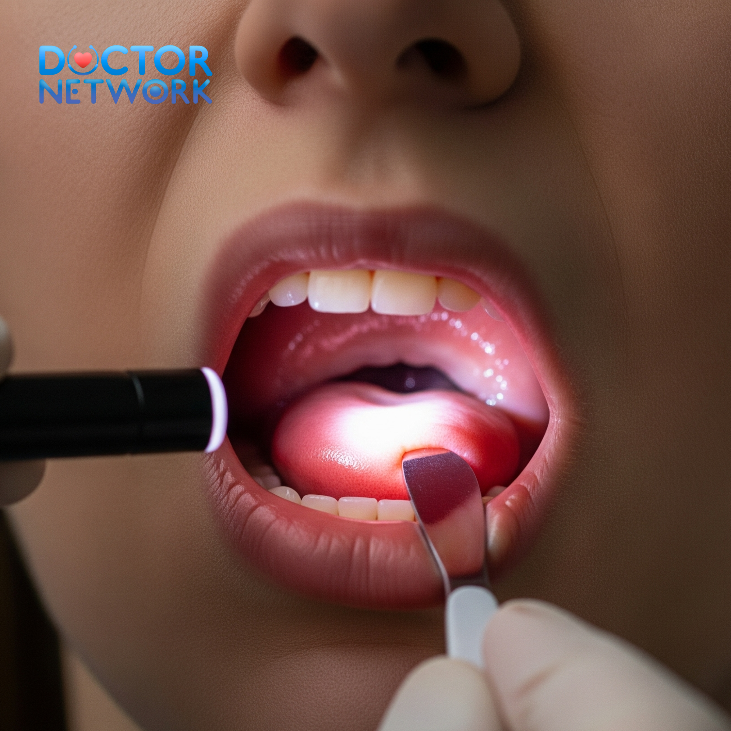
Odontogenic pain represents the most frequent misdiagnosis source for trigeminal neuralgia, leading to unnecessary dental procedures and delayed appropriate treatment. Dental pathologies produce continuous, throbbing, or pressure-like sensations distinctly different from TN’s sharp, electric shock-like quality. Pulpitis, periapical abscesses, and periodontal infections create persistent discomfort that worsens with biting pressure, temperature changes, or positional modifications.
Cracked tooth syndrome generates sharp, momentary pain during chewing or temperature exposure, but lacks the spontaneous, trigger-sensitive nature characteristic of trigeminal neuralgia. The pain localizes precisely to the affected tooth, responds to dental anesthesia, and demonstrates clear radiographic or clinical evidence of structural damage.
Atypical facial pain of dental origin may present with burning, aching, or cramping sensations following dental procedures, extraction complications, or nerve damage during oral surgery. This neuropathic dental pain differs from TN through its constant nature, burning quality, and direct relationship to previous dental interventions.
Diagnostic Differentiators:
- Location: Precise tooth localization vs. nerve distribution
- Quality: Throbbing/pressure vs. electric shock-like
- Duration: Continuous vs. brief episodes
- Triggers: Biting/temperature vs. light touch
- Response: Dental anesthesia effective vs. ineffective
Musculoskeletal and Joint Disorders
Temporomandibular joint disorders (TMJD) create complex orofacial pain patterns that frequently overlap with trigeminal neuralgia presentations. TMJ dysfunction produces deep, aching pain within the joint capsule, often radiating to the temporal region, ear, and neck. The pain typically correlates with jaw movement, exhibits diurnal variation, and associates with joint sounds, limited opening, or locking episodes.
Myofascial pain dysfunction syndrome (MPDS) affects masticatory muscles through chronic tension, bruxism, or postural abnormalities. Trigger points within the masseter, temporalis, and pterygoid muscles create referred pain patterns that may simulate trigeminal distributions. Unlike TN, myofascial pain responds to muscle palpation, demonstrates muscle tenderness, and improves with muscle relaxation techniques.
Cervical spine disorders contribute to facial pain through trigemino-cervical convergence mechanisms. Upper cervical joint dysfunction, particularly atlantooccipital and atlantoaxial pathology, can refer pain to trigeminal territories through shared neural pathways. Cervicogenic headache patterns may extend into facial regions, creating diagnostic confusion with trigeminal neuralgia.
| Condition | Pain Quality | Location | Triggers | Associated Features |
|---|---|---|---|---|
| TMJD | Deep, aching | Joint, temple | Jaw movement | Joint sounds, locking |
| MPDS | Dull, cramping | Muscle belly | Palpation | Muscle tenderness |
| Cervicogenic | Throbbing | Neck to face | Head movement | Neck stiffness |
Other Neuropathic Pains and Headaches
Glossopharyngeal neuralgia (GPN) shares striking similarities with trigeminal neuralgia but affects the ninth cranial nerve, producing pain in the throat, tongue base, and ear regions. GPN episodes occur during swallowing, speaking, or coughing, with pain radiating from the tonsillar area to the ear. The electric shock-like quality resembles TN, but anatomical distribution and swallowing triggers distinguish these conditions.
Postherpetic neuralgia (PHN) develops following herpes zoster infections affecting trigeminal nerve distributions, particularly the ophthalmic division. PHN creates constant burning, tingling, or allodynic sensations within the previous rash distribution. Unlike TN’s episodic nature, PHN produces persistent discomfort with hyperesthesia and allodynia to light touch.
Trigeminal neuropathy encompasses various non-neuralgic trigeminal nerve disorders characterized by continuous facial numbness, burning, or dysesthetic sensations. These conditions result from nerve damage, inflammation, or demyelination without the paroxysmal discharge patterns characteristic of classic trigeminal neuralgia. The pain quality differs significantly, presenting as constant burning, tingling, or pins-and-needles sensations rather than electric shock-like episodes.
Neuropathic Pain Comparison:
- Trigeminal Neuralgia: Electric, episodic, trigger-sensitive
- Glossopharyngeal Neuralgia: Electric, throat-based, swallowing triggers
- Postherpetic Neuralgia: Burning, constant, previous rash history
- Trigeminal Neuropathy: Burning, continuous, sensory changes
Headache Syndromes and Other Conditions
Sinus infections (sinusitis) produce deep, pressure-like facial pain that may simulate trigeminal neuralgia in anatomical distribution. Acute sinusitis creates constant, throbbing discomfort with nasal congestion, purulent discharge, and facial tenderness over affected sinuses. The pain typically worsens with bending forward, coughing, or sinus pressure, distinguishing it from TN’s trigger-sensitive, electric shock-like episodes.
Migraine headaches occasionally present with unilateral facial pain components, particularly in complex migraine variants or atypical presentations. Migrainous facial pain demonstrates typical migraine characteristics including photophobia, phonophobia, nausea, and throbbing quality lasting hours to days. The continuous nature and associated autonomic symptoms differentiate migraine from TN’s brief, electric shock-like episodes.
Cluster headaches create intense, unilateral orbital or temporal pain with characteristic autonomic features including lacrimation, nasal congestion, and Horner’s syndrome. The pain occurs in cyclical patterns with predictable timing, lasting 15 minutes to 3 hours, distinctly different from TN’s seconds-to-minutes episodes. Cluster headache pain focuses around the eye and temple rather than trigeminal nerve distributions.
Additional Mimicking Conditions:
- Giant Cell Arteritis: Temporal artery inflammation with jaw claudication
- Paroxysmal Hemicrania: Orbital pain with autonomic symptoms
- SUNCT/SUNA: Short-lasting unilateral neuralgiform attacks with conjunctival injection
- Atypical Facial Pain: Continuous burning without clear etiology
Diagnostic Approach: Differentiating TN from Mimics
Clinical Assessment and History Taking
Trigeminal neuralgia diagnosis relies fundamentally on comprehensive clinical evaluation, emphasizing detailed pain characterization and trigger identification. The diagnostic process begins with meticulous pain history documentation, focusing on onset patterns, pain quality descriptors, episode duration, frequency, and specific triggering stimuli that provoke attacks.
Clinicians must systematically explore pain characteristics using standardized descriptors to differentiate TN’s electric shock-like quality from other facial pain types. Patients typically describe TN pain as “lightning bolts,” “electrical shocks,” or “stabbing knife-like” sensations, distinctly different from the burning, throbbing, or pressure-like qualities of other conditions.
Trigger zone identification represents a crucial diagnostic component, as TN demonstrates characteristic light-touch sensitivity in specific facial regions. Common trigger zones include the nasolabial fold, corner of the mouth, and areas along the maxillary or mandibular nerve distributions. Gentle examination of these regions with cotton swabs or light finger pressure can reproduce typical pain episodes in genuine TN cases.
Essential History Components:
- Pain onset circumstances and progression
- Detailed pain quality descriptors
- Episode duration and frequency patterns
- Specific trigger identification
- Previous treatments and responses
- Associated neurological symptoms
- Family history of similar conditions
Physical Examination Techniques
Comprehensive physical examination in suspected trigeminal neuralgia cases focuses on neurological assessment while systematically excluding other facial pain sources. The cranial nerve examination emphasizes trigeminal nerve function evaluation across all three divisions, assessing light touch, pinprick sensation, and corneal reflex responses.
Sensory examination utilizes standardized techniques including cotton swab light touch, pinprick testing, and vibration assessment across trigeminal dermatomes. Normal sensory function supports classic TN diagnosis, while sensory deficits suggest secondary causes or alternative diagnoses requiring further investigation.
Trigger zone mapping involves systematic light stimulation of suspected trigger areas using cotton swabs, gentle finger pressure, or soft brushes. Positive trigger responses reproduce characteristic pain episodes, confirming TN diagnosis. The examination must proceed cautiously to avoid excessive patient discomfort while gathering essential diagnostic information.
Motor function assessment evaluates masseter and temporalis muscle strength, jaw opening symmetry, and lateral excursion capabilities. Weakness or asymmetry suggests structural pathology requiring advanced imaging evaluation. Temporomandibular joint examination includes palpation, range of motion testing, and auscultation for joint sounds.
Diagnostic Imaging and Tests
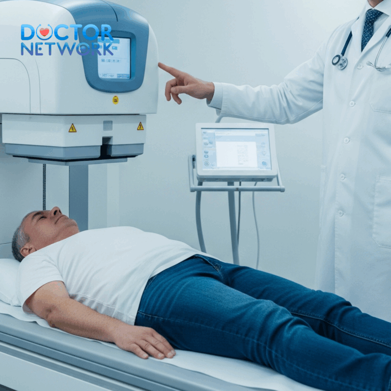
Magnetic Resonance Imaging (MRI) serves as the gold standard for trigeminal neuralgia evaluation, providing detailed visualization of trigeminal nerve anatomy and surrounding structures. High-resolution, thin-section sequences optimize nerve visualization and neurovascular relationship assessment. Specialized sequences including FIESTA (Fast Imaging Employing Steady-state Acquisition) or CISS (Constructive Interference in Steady State) enhance nerve-vessel interface delineation.
MRI evaluation focuses on identifying neurovascular compression at the trigeminal nerve root entry zone, assessing for vessel displacement, nerve distortion, or atrophy. Secondary causes including multiple sclerosis plaques, tumors, or structural abnormalities require systematic evaluation throughout the brainstem and posterior fossa regions.
Magnetic Resonance Angiography (MRA) complements standard MRI by providing detailed vascular imaging, identifying aberrant vessels, aneurysms, or vascular malformations contributing to nerve compression. Time-of-flight or contrast-enhanced sequences optimize arterial visualization and flow assessment.
Advanced imaging techniques include diffusion tensor imaging (DTI) for nerve microstructure evaluation and functional MRI sequences assessing trigeminal nerve function. These specialized techniques provide additional diagnostic information in complex cases where standard imaging appears normal.
Imaging Protocol Components:
- High-resolution T1 and T2-weighted sequences
- FIESTA or CISS nerve-specific sequences
- Post-contrast T1-weighted images
- MRA vascular evaluation
- Dedicated trigeminal nerve imaging
Role of Neurological Evaluation
Comprehensive neurological evaluation provides essential diagnostic information while excluding secondary causes of trigeminal neuralgia. The assessment encompasses detailed neurological history, systematic cranial nerve examination, and comprehensive sensory testing across trigeminal distributions.
Specialized testing includes trigeminal reflex evaluation, assessing both the masseter reflex and blink reflex responses. Abnormal reflex patterns suggest structural pathology or secondary TN causes requiring further investigation. Corneal reflex testing evaluates trigeminal nerve ophthalmic division function, with absent responses indicating significant nerve dysfunction.
Quantitative sensory testing (QST) provides objective sensory function assessment using standardized thermal and mechanical stimuli. QST results help differentiate classic TN from neuropathic conditions and monitor treatment responses over time. Neurophysiological testing may include nerve conduction studies or electromyography in selected cases.
Utilizing Functional Medicine Tests
Functional medicine approaches complement traditional diagnostic methods by identifying systemic factors contributing to neurological dysfunction and pain amplification. These comprehensive assessments evaluate inflammation markers, hormonal balance, gastrointestinal health, and nutritional status that may influence trigeminal neuralgia severity and treatment response.
C-Reactive Protein (CRP) testing measures systemic inflammation levels that can exacerbate neuropathic pain conditions. Elevated inflammatory markers suggest underlying immune activation that may contribute to nerve sensitization and pain amplification. Anti-inflammatory interventions targeting elevated CRP levels may provide adjunctive therapeutic benefits.
The DUTCH Complete hormonal assessment evaluates comprehensive hormone profiles including cortisol patterns, sex hormones, and neurotransmitter metabolites. Cortisol dysregulation affects pain perception, stress response, and nervous system function. Identifying hormonal imbalances enables targeted interventions that may reduce TN symptom severity.
Gastrointestinal health evaluation through comprehensive stool analysis (GI-MAP) assesses microbiome composition, inflammatory markers, and digestive function. Gut-brain axis dysfunction contributes to systemic inflammation and neurological symptoms. Optimizing gastrointestinal health through targeted interventions may reduce overall inflammatory burden and improve neurological function.
Functional Medicine Test Panel:
- Inflammation: CRP, ESR, cytokine profiles
- Hormonal: DUTCH Complete, thyroid function
- Gastrointestinal: GI-MAP, food sensitivity testing
- Nutritional: Vitamin B12, folate, vitamin D levels
- Metabolic: Homocysteine, methylation status
Treatment Options for Trigeminal Neuralgia
Medications
Pharmacological management represents the first-line treatment approach for trigeminal neuralgia, with antiepileptic drugs demonstrating superior efficacy compared to traditional analgesics. Carbamazepine remains the gold standard medication, providing effective pain control in approximately 80% of patients with classic TN. The drug stabilizes sodium channels, reducing aberrant nerve firing and paroxysmal discharge patterns characteristic of TN.
Oxcarbazepine serves as an alternative first-line option with potentially fewer side effects than carbamazepine while maintaining similar efficacy. The medication offers improved tolerability profiles with reduced drug interactions and monitoring requirements. Dosage optimization requires gradual titration to balance therapeutic efficacy with acceptable side effect profiles.
Second-line medications include gabapentin, pregabalin, and baclofen, either as monotherapy or combination treatments for refractory cases. Gabapentin and pregabalin provide neuropathic pain relief through calcium channel modulation, while baclofen offers muscle relaxation and GABA-ergic effects. Combination therapy may enhance efficacy when single agents provide insufficient relief.
Medication Efficacy Comparison:
| Medication | Efficacy Rate | Common Side Effects | Monitoring Requirements |
|---|---|---|---|
| Carbamazepine | 80-90% | Drowsiness, dizziness, nausea | CBC, liver function, sodium |
| Oxcarbazepine | 75-85% | Fatigue, headache, nausea | Sodium levels |
| Gabapentin | 60-70% | Sedation, weight gain | Minimal |
| Pregabalin | 65-75% | Dizziness, peripheral edema | Minimal |
| Baclofen | 50-60% | Weakness, confusion | Gradual withdrawal required |
Surgical Procedures
Surgical intervention becomes necessary when medications fail to provide adequate pain control or produce intolerable side effects limiting quality of life. Multiple surgical options exist, each with distinct mechanisms, efficacy rates, and risk profiles requiring careful patient selection and counseling.
Microvascular decompression (MVD) represents the most definitive surgical treatment for classic TN caused by neurovascular compression. The procedure involves posterior fossa craniotomy with microsurgical identification and separation of compressing vessels from the trigeminal nerve root. MVD provides excellent long-term pain relief in 85-95% of patients with low recurrence rates.
Percutaneous procedures offer less invasive alternatives for patients unsuitable for open surgery or preferring minimal intervention approaches. Glycerol rhizotomy involves stereotactic injection of glycerol into Meckel’s cave, creating controlled nerve damage to reduce pain transmission. Balloon compression utilizes controlled compression of the trigeminal ganglion to achieve pain relief.
Stereotactic radiosurgery, including Gamma Knife and CyberKnife systems, delivers focused radiation to the trigeminal nerve root, creating controlled lesions that reduce pain transmission. This non-invasive approach avoids surgical risks while providing effective pain relief in 70-85% of patients. Treatment effects develop gradually over weeks to months following radiation delivery.
Surgical Options Summary:
- Microvascular Decompression: Highest efficacy, preserves nerve function
- Glycerol Rhizotomy: Minimally invasive, repeatable procedure
- Balloon Compression: Rapid onset, predictable sensory loss
- Radiosurgery: Non-invasive, delayed onset, excellent for elderly patients
Non-Invasive Therapies
Complementary and alternative therapies provide valuable adjunctive treatments for trigeminal neuralgia management, particularly when combined with conventional medical approaches. These interventions focus on pain modulation, stress reduction, and overall quality of life improvement while avoiding additional medication side effects.
Acupuncture demonstrates promising results in TN management through endorphin release, nerve modulation, and anti-inflammatory effects. Systematic studies report significant pain reduction and improved quality of life measures in TN patients receiving regular acupuncture treatments. Treatment protocols typically involve multiple sessions targeting specific acupoints related to facial pain and nervous system function.
Physical therapy interventions include gentle facial exercises, posture correction, and cervical spine mobilization techniques. Upper cervical dysfunction may contribute to TN symptoms through trigemino-cervical convergence mechanisms. Addressing cervical spine alignment and muscle tension may provide additional pain relief and prevent trigger zone sensitivity.
Nerve blocks using local anesthetics provide temporary pain relief while confirming diagnosis and predicting surgical outcomes. Trigeminal nerve blocks target specific divisions affected by pain, offering both therapeutic and diagnostic value. These procedures help differentiate TN from other facial pain conditions while providing temporary symptom relief.
Chiropractic Care
Upper cervical chiropractic care addresses potential cervical spine contributions to trigeminal neuralgia through precise spinal alignment correction and nervous system optimization. Specialized techniques including NUCCA (National Upper Cervical Chiropractic Association) and Toggle Recoil procedures focus on atlas and axis alignment restoration, potentially reducing nerve irritation and improving neurological function.
The upper cervical spine’s intimate relationship with brainstem structures and cranial nerve nuclei suggests that cervical misalignments may contribute to trigeminal nerve dysfunction. Careful analysis of cervical spine alignment using advanced imaging and postural assessment identifies specific misalignment patterns requiring correction.
Treatment protocols involve gentle, precise adjustments designed to restore normal cervical spine biomechanics without aggressive manipulation. These techniques emphasize neurological function improvement rather than forceful joint manipulation, making them suitable for sensitive TN patients who may not tolerate traditional chiropractic approaches.
Upper Cervical Benefits:
- Improved nerve function through spinal alignment
- Reduced muscle tension and trigger point activity
- Enhanced blood flow to neural structures
- Decreased inflammatory responses
- Natural pain relief without medications
Living with Trigeminal Neuralgia & Coping Strategies
Lifestyle Adjustments
Successful trigeminal neuralgia management requires comprehensive lifestyle modifications designed to minimize trigger exposure while maintaining quality of life and daily functioning. These adaptations focus on identifying and avoiding specific triggers while developing alternative approaches to essential activities.
Dietary modifications play a crucial role in TN management, emphasizing foods that minimize chewing requirements and avoid temperature extremes. Soft, room-temperature foods reduce mechanical stimulation while providing adequate nutrition. Food preparation techniques including chopping, blending, or cooking until soft consistency help maintain dietary variety while accommodating TN limitations.
Environmental modifications protect against weather-related triggers including cold air, wind, and barometric pressure changes. Protective clothing including scarves, face masks, or windbreakers help shield trigger zones from environmental stimuli. Indoor climate control maintains consistent temperature and humidity levels, reducing sudden environmental changes that may provoke attacks.
Personal care routines require careful adaptation to minimize facial stimulation while maintaining hygiene and appearance. Gentle techniques for tooth brushing, face washing, and cosmetic application help preserve personal care standards without triggering pain episodes. Electric toothbrushes set on gentle settings may provide effective cleaning with reduced manual pressure.
Daily Living Adaptations:
| Activity | Challenge | Adaptation Strategy |
|---|---|---|
| Eating | Chewing triggers pain | Soft foods, small bites, room temperature |
| Speaking | Facial movement triggers | Minimize talking, written communication |
| Dental care | Direct touch sensitivity | Soft brushes, gentle technique, electric brushes |
| Face washing | Water contact triggers | Gentle patting, avoid rubbing motions |
| Cold weather | Environmental triggers | Protective clothing, warm environments |
Stress Management
Stress represents a significant trigger factor for trigeminal neuralgia episodes, creating a cyclical pattern where pain increases stress levels, which subsequently trigger additional pain episodes. Effective stress management strategies break this cycle while improving overall pain tolerance and quality of life.
Mindfulness meditation and deep breathing exercises provide immediate stress relief while developing long-term resilience against pain-related anxiety. Regular meditation practice helps patients develop greater awareness of stress responses and early intervention strategies to prevent escalation into full pain episodes.
Progressive muscle relaxation techniques specifically benefit TN patients by reducing facial and cervical muscle tension that may contribute to trigger zone sensitivity. Systematic relaxation of facial muscles, jaw, and neck regions helps decrease overall tension levels while providing direct therapeutic benefits for pain management.
Cognitive-behavioral therapy (CBT) addresses the psychological aspects of chronic pain, helping patients develop coping strategies, challenge negative thought patterns, and maintain functional capacity despite ongoing symptoms. CBT techniques prove particularly valuable for managing anticipatory anxiety and fear avoidance behaviors common in TN patients.
Support Systems
Comprehensive support systems provide essential emotional and practical assistance for individuals managing trigeminal neuralgia’s complex challenges. These networks encompass family members, healthcare providers, support groups, and community resources that collectively enhance coping capacity and treatment outcomes.
Family education and involvement ensures that loved ones understand TN’s unpredictable nature, trigger sensitivity, and impact on daily functioning. Educated family members can provide appropriate support during pain episodes while avoiding well-intentioned actions that might trigger attacks.
Professional support groups, both in-person and online, connect TN patients with others facing similar challenges. These communities provide valuable information sharing, emotional support, and practical advice from individuals with lived experience managing TN symptoms.
Support Network Components:
- Medical Team: Neurologists, pain specialists, primary care providers
- Family/Friends: Educated support persons, emergency contacts
- Support Groups: TN associations, online communities, local groups
- Professional Services: Mental health counselors, social workers
- Community Resources: Transportation, meal assistance, household help
Healthcare provider coordination ensures comprehensive care delivery across multiple specialists and treatment modalities. Regular communication between neurologists, primary care physicians, and other healthcare providers optimizes treatment outcomes while preventing conflicting interventions or medication interactions.
Challenges in Diagnosis and Referral Considerations
Diagnostic Complexity and Common Pitfalls
Trigeminal neuralgia diagnosis presents significant challenges due to its symptom overlap with numerous facial pain conditions, leading to frequent misdiagnosis and inappropriate treatment delays. The complexity stems from TN’s reliance on subjective pain descriptions, variable presentation patterns, and the absence of objective diagnostic markers that definitively confirm the condition.
Primary care physicians face particular challenges in TN diagnosis due to limited exposure to this relatively rare condition and the need for specialized neurological assessment tools. The average delay from symptom onset to accurate diagnosis ranges from 6 months to several years, during which patients may undergo unnecessary dental procedures, surgical interventions, or inappropriate medication trials.
Common diagnostic errors include attributing TN symptoms to dental pathology, leading to unnecessary tooth extractions, root canal procedures, or oral surgeries that fail to address the underlying neurological condition. These interventions not only prove ineffective but may actually worsen symptoms through additional tissue trauma and nerve irritation.
Diagnostic Red Flags Suggesting Incorrect TN Diagnosis:
- Continuous rather than episodic pain patterns
- Bilateral facial pain distribution
- Absence of trigger zone sensitivity
- Pain not responding to carbamazepine
- Associated sensory deficits or motor weakness
- Pain triggered by deep pressure rather than light touch
Referral Guidelines and Specialist Consultation
Appropriate referral to neurological specialists becomes essential when primary care assessment cannot confidently establish TN diagnosis or when initial treatment approaches fail to provide adequate symptom control. Early specialist consultation prevents unnecessary interventions while ensuring access to advanced diagnostic techniques and specialized treatment options.
Neurologists possess specialized expertise in cranial nerve disorders, advanced imaging interpretation, and complex medication management required for optimal TN treatment. Their evaluation includes sophisticated neurological assessment techniques, specialized diagnostic testing, and access to advanced treatment modalities unavailable in primary care settings.
Pain management specialists offer additional expertise in interventional procedures, complex medication regimens, and multidisciplinary treatment approaches for refractory cases. These specialists provide access to nerve blocks, neuromodulation techniques, and comprehensive pain management strategies when standard treatments prove insufficient.
Referral Indications:
- Uncertain TN diagnosis despite thorough evaluation
- Failure to respond to first-line medication therapy
- Intolerable medication side effects
- Bilateral or atypical pain presentations
- Associated neurological symptoms or deficits
- Patient preference for specialized care
Neurosurgical consultation becomes necessary for patients considering surgical intervention due to medication failure, intolerable side effects, or personal preference for definitive treatment. Neurosurgeons specializing in functional neurosurgery provide expertise in microvascular decompression, stereotactic radiosurgery, and percutaneous procedures tailored to individual patient needs and risk profiles.
5 frequently asked questions about “what can be mistaken for trigeminal neuralgia”
1. What conditions can cause facial pain that mimics trigeminal neuralgia?
Several conditions can cause facial pain similar to TN, including dental problems (such as tooth infections or abscesses), headache syndromes like cluster headaches, temporal arteritis, sinusitis, tumors in the brain or face, temporomandibular joint (TMJ) dysfunction, postherpetic neuralgia (nerve pain after shingles), myofascial pain dysfunction syndrome, and glossopharyngeal neuralgia.
2. How can dental pain be distinguished from trigeminal neuralgia?
Dental pain usually presents as a more continuous, localized pain often triggered or relieved by eating, temperature changes, or biting. It often resolves quickly after dental treatment or removal of the stimulus (like cold food). In contrast, TN pain is typically sudden, sharp, electric shock-like, brief, and triggered by innocuous stimuli such as talking or light touch. TN pain does not fade quickly after such triggers and is not usually associated with visible dental abnormalities.
3. Can headache disorders be confused with trigeminal neuralgia?
Yes. Cluster headaches and other short-lasting unilateral neuralgiform headache attacks (such as SUNA and SUNCT) can cause severe, unilateral facial or orbital pain that resembles TN. However, cluster headaches tend to last longer and are often accompanied by autonomic symptoms like tearing or nasal congestion. Migraines and paroxysmal hemicrania also cause facial or head pain but usually have additional features such as photophobia, phonophobia, or longer-lasting pain episodes.
4. What role do nerve-related conditions play in mimicking trigeminal neuralgia?
Postherpetic neuralgia, glossopharyngeal neuralgia, and trigeminal neuropathy can present with facial pain similar to TN. Postherpetic neuralgia usually follows a shingles rash and causes continuous burning pain, unlike the intermittent shocks of TN. Glossopharyngeal neuralgia causes pain in the throat, tongue, or ear triggered by swallowing or talking. Trigeminal neuropathy causes more constant, burning, or pins-and-needles sensations rather than sharp stabbing pain.
5. How is trigeminal neuralgia accurately diagnosed to avoid confusion with other conditions?
Diagnosis relies on a thorough clinical history and physical examination focusing on the characteristic pain pattern: recurrent, brief, unilateral, stabbing or shock-like pain in the trigeminal nerve distribution triggered by light touch or movement. Examination helps exclude dental, TMJ, or sinus problems. Imaging, especially MRI, is used to rule out secondary causes like tumors or multiple sclerosis. Differentiating TN from other facial pains requires careful assessment of pain quality, triggers, associated symptoms, and medical history.
Conclusion: Towards Accurate Diagnosis and Improved Quality of Life
Effective trigeminal neuralgia diagnosis and management requires a comprehensive, multidisciplinary approach that integrates detailed clinical assessment, advanced diagnostic techniques, and personalized treatment strategies. Healthcare providers must develop sophisticated understanding of TN’s complex presentation patterns while maintaining awareness of numerous mimicking conditions that can lead to diagnostic confusion and inappropriate treatment.
The integration of traditional neurological evaluation with advanced imaging techniques, functional medicine assessments, and specialized diagnostic procedures enhances diagnostic accuracy while reducing the risk of misdiagnosis and treatment delays. This comprehensive approach ensures that patients receive appropriate interventions tailored to their specific condition and individual needs.
Accurate TN diagnosis serves as the foundation for effective treatment planning, enabling healthcare providers to select optimal therapeutic approaches that maximize pain relief while minimizing side effects and complications. Early diagnosis and appropriate treatment significantly improve patient outcomes, quality of life, and long-term prognosis.
The complexity of facial pain diagnosis demands continued medical education, interdisciplinary collaboration, and patient advocacy to ensure that individuals suffering from trigeminal neuralgia receive prompt, accurate diagnosis and effective treatment. Specialized clinics focusing on facial pain disorders provide essential resources for complex cases requiring expert evaluation and management.
Through continued research, improved diagnostic techniques, and enhanced treatment options, the future holds promise for better outcomes and improved quality of life for individuals affected by trigeminal neuralgia and related facial pain conditions. The key lies in maintaining high clinical suspicion, utilizing appropriate diagnostic resources, and ensuring timely access to specialized care when needed.
References
1. Dental and Odontogenic Pathologies
This is one of the most common sources of misdiagnosis. Patients often see a dentist first, and unnecessary dental procedures may be performed before a neurological cause is considered.
Why it’s mistaken: Pain from conditions like a cracked tooth, pulpitis (inflammation of the tooth’s pulp), or a dental abscess can be sharp, severe, and radiate along the jaw, mimicking the distribution of the trigeminal nerve.
Evidence/Study 1:
Title: Odontogenic Pain Mimicking Trigeminal Neuralgia: A Diagnostic Challenge.
Author(s): Melis M, Secci S.
Source: Cephalalgia, 2007.
Link: https://journals.sagepub.com/doi/abs/10.1111/j.1468-2982.2007.01373.x
Key Findings: The study highlights cases where patients were initially diagnosed with TN but were later found to have underlying dental issues. It emphasizes that a thorough dental examination, including radiographs and percussion tests, is essential to rule out odontogenic causes before diagnosing idiopathic TN.
Evidence/Study 2:
Title: When to suspect a non-odontogenic origin for toothache.
Author(s): Graff-Radford SB, Solberg WK.
Source: Mouth, 1992. (A foundational paper often cited in this context).
Key Findings: This article outlines clinical clues that suggest a non-dental cause for tooth pain. Pain that is not affected by thermal stimuli (hot/cold), is spontaneous, or crosses the midline of the face is less likely to be of dental origin and may point towards a neuropathic condition like TN.
2. Temporomandibular Disorders (TMD)
TMD involves problems with the jaw joint and the muscles of mastication (chewing).
Why it’s mistaken: Pain is located in the preauricular area (in front of the ear), jaw, and face. It can be triggered by chewing, talking, or yawning, which can also be triggers for TN.
Evidence/Study:
Title: Differential diagnosis of temporomandibular disorders and other orofacial pain conditions.
Author(s): Glaros AG, Williams K, Laustrup D.
Source: A Textbook of Occlusion, 2018 (Chapter). A good overview is also in the Journal of Oral & Facial Pain and Headache.
Link: (This is a chapter, but a representative article on the topic is): https://www.quintpub.com/journals/ofph/abstract.php?article_id=19039
Key Findings: The key differentiator is the quality of pain. TMD pain is typically a dull, aching, and continuous discomfort, often associated with joint sounds (clicking, popping) and limited jaw movement. In contrast, classic TN pain is paroxysmal, sharp, and electrical. However, atypical facial pain and TMD can overlap significantly.
3. Postherpetic Neuralgia (PHN)
PHN is a complication of shingles (herpes zoster infection) that affects a nerve.
Why it’s mistaken: If the shingles outbreak affects the ophthalmic division of the trigeminal nerve, it can result in persistent facial pain in the same distribution as TN.
Evidence/Study:
Title: Postherpetic Neuralgia: A Review.
Author(s): Johnson RW, Rice AS.
Source: New England Journal of Medicine, 2014.
Key Findings: A key diagnostic clue for PHN is the patient’s history. There will be a preceding shingles rash in the painful area. The pain character is also often different—typically described as a constant burning, aching, or itching, although it can have superimposed sharp, stabbing pains similar to TN.
4. Multiple Sclerosis (MS)
TN can be a symptom of MS, known as “symptomatic trigeminal neuralgia.”
Why it’s mistaken: The pain presentation is identical to classic TN. However, the underlying cause is a demyelinating plaque in the brainstem at the trigeminal nerve root entry zone, not vascular compression.
Evidence/Study:
Title: Trigeminal neuralgia in multiple sclerosis: a new physiopathological hypothesis.
Author(s): Cruccu G, Biasiotta A, Di Stefano G, et al.
Source: Neurology, 2016.
Key Findings: This study discusses the mechanisms of TN in MS. The presence of TN in a younger patient (under 40), bilateral symptoms, or associated sensory loss should raise high suspicion for MS. An MRI is crucial for diagnosis to look for demyelinating plaques.
5. Trigeminal Autonomic Cephalalgias (TACs)
This group of headache disorders includes Cluster Headache and Paroxysmal Hemicrania.
Why it’s mistaken: The pain is severe, unilateral, and located in the trigeminal distribution (often around the eye).
Evidence/Study:
Title: The Trigeminal Autonomic Cephalalgias: Diagnosis and Management.
Author(s): Wei DY, Goadsby PJ.
Source: Current Pain and Headache Reports, 2021.
Link: https://link.springer.com/article/10.1007/s11916-021-00976-y
Key Findings: The defining feature of TACs is the presence of prominent cranial autonomic symptoms on the same side as the pain. These include conjunctival injection (red eye), lacrimation (tearing), nasal congestion, rhinorrhea (runny nose), eyelid edema, and/or a drooping eyelid (ptosis). These autonomic features are typically absent or minimal in classic TN. The duration and frequency of attacks also differ.
6. Persistent Idiopathic Facial Pain (PIFP)
Formerly known as Atypical Facial Pain, this is a diagnosis of exclusion.
Why it’s mistaken: It causes persistent facial pain without a clear organic cause.
Evidence/Study:
Title: Persistent idiopathic facial pain.
Author(s): Forssell H, Jääskeläinen S, List T, et al.
Source: Cephalalgia, 2020 (as part of the International Classification of Orofacial Pain, ICOP).
Link: https://journals.sagepub.com/doi/full/10.1177/0333102419893827
Key Findings: PIFP is characterized by daily, persistent facial pain that is poorly localized and does not follow a specific nerve distribution. The pain is often described as dull, aching, or burning, and lacks the paroxysmal, electric-shock quality of classic TN. It is not associated with sensory loss or other physical signs.
7. Other Less Common Mimics
Sinusitis: Inflammation of the paranasal sinuses can cause pressure and pain over the cheeks and forehead. The pain is usually dull, constant, and associated with other sinus symptoms.
Giant Cell Arteritis (Temporal Arteritis): An inflammation of the arteries, typically in older adults (>50). It can cause jaw claudication (pain with chewing) and headache. A key differentiator is often an elevated erythrocyte sedimentation rate (ESR) on a blood test.
Tumors or Cysts: A tumor (like an acoustic neuroma or meningioma) in the cerebellopontine angle can compress the trigeminal nerve, causing symptomatic TN. An MRI is essential to rule this out.
Kiểm Duyệt Nội Dung
More than 10 years of marketing communications experience in the medical and health field.
Successfully deployed marketing communication activities, content development and social networking channels for hospital partners, clinics, doctors and medical professionals across the country.
More than 6 years of experience in organizing and producing leading prestigious medical programs in Vietnam, in collaboration with Ho Chi Minh City Television (HTV). Typical programs include Nhật Ký Blouse Trắng, Bác Sĩ Nói Gì, Alo Bác Sĩ Nghe, Nhật Ký Hạnh Phúc, Vui Khỏe Cùng Con, Bác Sỹ Mẹ, v.v.
Comprehensive cooperation with hundreds of hospitals and clinics, thousands of doctors and medical experts to join hands in building a medical content and service platform on the Doctor Network application.