


Magnetic Resonance Imaging (MRI) scans with contrast agents represent one of modern medicine’s most powerful diagnostic tools, providing unprecedented clarity in detecting abnormalities, tumors, and vascular conditions. The gadolinium-based contrast dye injected intravenously enhances tissue visibility, allowing radiologists to distinguish between healthy and diseased tissue with remarkable precision. However, patients frequently encounter a crucial pre-procedure requirement: fasting before their contrast-enhanced MRI examination.
“Why do you have to fast before MRI with contrast” – This comprehensive guide explores the medical necessity behind pre-scan fasting, examining both patient safety protocols and image quality optimization. We’ll delve into specific scanning procedures that mandate dietary restrictions, appropriate fasting durations for different examinations, and detailed preparation guidelines for various patient populations. Understanding these requirements ensures optimal diagnostic outcomes while maintaining the highest safety standards during your medical imaging procedure.
Understanding MRI with Contrast Enhancement
Contrast-enhanced magnetic resonance imaging involves the intravenous administration of gadolinium-based contrast agents, typically injected through an IV line established in the patient’s arm or hand. These paramagnetic substances dramatically improve visualization of soft tissues, blood vessels, and pathological processes that might remain invisible on standard MRI sequences.
The contrast material functions by altering the magnetic properties of surrounding tissues, creating enhanced signal intensity on T1-weighted images. This enhancement proves particularly valuable when diagnosing complex conditions including malignant neoplasms, multiple sclerosis lesions, cardiac pathology, infectious processes, and inflammatory diseases. Radiologists rely on contrast enhancement patterns to differentiate between various tissue types and identify subtle abnormalities that could indicate serious medical conditions.
Modern gadolinium compounds demonstrate excellent safety profiles for most patients, though they require careful consideration in individuals with compromised renal function. The contrast agent circulates through the vascular system before being filtered and eliminated by the kidneys, typically within 24-48 hours post-injection.
Common clinical applications include:
- Oncological staging and tumor characterization
- Neurological disorder assessment (multiple sclerosis, brain tumors)
- Cardiovascular evaluation (myocardial infarction, cardiomyopathy)
- Musculoskeletal imaging (joint pathology, soft tissue masses)
- Abdominal organ assessment (liver lesions, pancreatic disorders)
The Essential Medical Reasons Behind Pre-Scan Fasting
Primary Safety Consideration: Preventing Aspiration Pneumonia
Fasting requirements exist primarily to eliminate life-threatening complications related to contrast agent administration and the supine positioning required during MRI examinations. Gadolinium-based contrast materials can trigger gastrointestinal side effects including nausea, vomiting, and gastric distress in susceptible patients.
When patients experience emesis while lying flat in the MRI scanner, the risk of pulmonary aspiration increases dramatically. Aspirated gastric contents can cause severe respiratory complications, including chemical pneumonitis, airway obstruction, and potentially fatal aspiration pneumonia. An empty stomach significantly reduces both the volume and acidity of potential aspirate, thereby minimizing these serious medical risks.
This safety consideration becomes even more critical when sedation or general anesthesia is required for claustrophobic patients, pediatric examinations, or complex interventional procedures. Anesthetic agents further suppress protective airway reflexes, making pre-procedure fasting an absolute medical necessity rather than a mere precautionary measure.
Secondary Benefit: Optimizing Diagnostic Image Quality
Fasting also enhances image quality by reducing motion artifacts caused by normal digestive processes. Food consumption stimulates peristaltic contractions throughout the gastrointestinal tract, creating movement that can blur MRI images and potentially obscure pathological findings.
These involuntary muscle contractions generate motion artifacts that appear as streaking or ghosting on the final images, particularly problematic when scanning areas adjacent to the digestive system. For abdominal and pelvic MRI examinations, eliminating peristalsis through fasting ensures optimal visualization of organs including the liver, pancreas, kidneys, and reproductive structures.
Additionally, fasting promotes gallbladder distension by allowing bile accumulation, improving visualization of the biliary system and facilitating detection of gallstones, polyps, or inflammatory changes. For specialized procedures like Magnetic Resonance Cholangiopancreatography (MRCP), an empty gastrointestinal tract eliminates fluid interference that could compromise biliary and pancreatic duct imaging.
Specific MRI Examinations Requiring Fasting Protocols
Different MRI procedures have varying fasting requirements based on anatomical regions being examined and the potential for contrast-related complications. Understanding these specific requirements helps patients prepare appropriately for their scheduled examinations.
| MRI Examination Type | Fasting Duration | Primary Reason | Special Considerations |
|---|---|---|---|
| Abdominal MRI with Contrast | 6-8 hours | Motion artifact reduction + Safety | Avoid fatty foods 24 hours prior |
| Pelvic MRI with Contrast | 4-6 hours | Safety + Organ visualization | May require bladder filling |
| MRCP (Biliary System) | 6-8 hours | Gallbladder distension + Image clarity | Water may be permitted |
| Cardiac MRI with Contrast | 2 hours food, 24 hours caffeine | Heart rate stabilization + Safety | No coffee, tea, chocolate, or stimulants |
| MRI-Guided Procedures | 8 hours | Anesthesia safety requirements | Complete NPO status required |
Abdominal and Pelvic MRI Examinations
Abdominal contrast-enhanced MRI requires fasting periods ranging from 6-8 hours to ensure optimal visualization of hepatic, pancreatic, and gastrointestinal structures. The empty stomach eliminates peristaltic motion artifacts while reducing the risk of contrast-induced nausea in this patient population.
Pelvic MRI examinations focus on reproductive organs, bladder, and surrounding soft tissues. Fasting helps minimize bowel gas and peristaltic motion that could obscure pathological findings in the pelvis. Some protocols may require controlled bladder filling to improve visualization of pelvic structures.
Magnetic Resonance Cholangiopancreatography (MRCP)
MRCP represents a specialized technique for evaluating the biliary system and pancreatic ducts without invasive procedures. This examination requires extended fasting periods (6-8 hours) to promote maximal gallbladder distension and eliminate gastric fluid that could interfere with biliary visualization.
The prolonged fasting allows bile concentration within the gallbladder, creating enhanced contrast against surrounding tissues and improving detection of choledocholithiasis, biliary strictures, or pancreatic duct abnormalities.
Cardiac MRI with Specialized Restrictions
Cardiac MRI examinations have unique fasting requirements due to the need for heart rate stabilization and rhythm control. While solid food restrictions are typically limited to 2 hours pre-scan, patients must avoid all caffeine-containing substances for 24 hours before examination.
This extended caffeine restriction includes coffee (regular and decaffeinated), tea, chocolate, energy drinks, and certain medications containing stimulants. Caffeine can affect cardiac rhythm and contractility, potentially interfering with functional cardiac assessment and pharmacological stress testing.

Fasting Duration Guidelines and Protocol Variations
Standard Fasting Periods by Examination Type
Fasting requirements vary significantly based on the specific MRI procedure, institutional protocols, and individual patient factors. Most contrast-enhanced examinations require fasting periods ranging from 4-8 hours, with timing calculated from the last oral intake to the scheduled scan time.
General fasting guidelines include:
- 4-6 hour fast: Standard for most contrast-enhanced MRI examinations
- 6-8 hour fast: Required for abdominal, pelvic, and biliary system imaging
- 8+ hour fast: Mandatory for procedures requiring sedation or general anesthesia
- Extended restrictions: Cardiac MRI with 24-hour caffeine avoidance
Emergency situations may necessitate contrast-enhanced imaging without adequate fasting time. In these cases, medical teams implement additional safety measures including gastric decompression, enhanced monitoring, and modified positioning to minimize aspiration risks.
Institutional Protocol Variations
Different medical facilities may have varying fasting protocols based on their clinical experience, patient population, and safety guidelines. Academic medical centers often implement longer fasting periods compared to outpatient imaging centers, reflecting their higher-risk patient populations and complex procedure requirements.
Some institutions allow clear liquids up to 2-3 hours before examination, while others require complete NPO (nothing by mouth) status. These variations reflect different risk assessments and safety philosophies rather than contradictory medical evidence.
Comprehensive Pre-Scan Preparation Guidelines
Essential First Steps and Medical History Documentation
Successful MRI preparation begins with thorough communication between patients and their healthcare team. Contact your physician’s office or the radiology department 24-48 hours before your scheduled examination to confirm specific fasting requirements and address any concerns about medication management during the fasting period.
Provide complete medical history information including previous allergic reactions (medications, contrast agents, food allergies), significant medical conditions (kidney disease, diabetes, heart conditions), prior surgical procedures, and current pregnancy status. This information helps the medical team assess contrast safety and determine appropriate monitoring requirements.
For female patients of childbearing age, pregnancy testing may be required before contrast administration. While MRI itself doesn’t use ionizing radiation, gadolinium contrast agents can cross the placental barrier and are generally avoided during pregnancy unless absolutely medically necessary.
Medication Management During Fasting
Most patients can continue their regular medications during fasting periods, taking pills with small sips of water as needed. However, medications normally taken with food may require timing adjustments or temporary modifications to prevent gastric irritation.
Special medication considerations:
- Diabetic patients: Require careful blood glucose monitoring and possible insulin adjustments
- Blood pressure medications: Usually continued with minimal water
- Heart medications: Typically maintained on normal schedule
- Blood thinners: May require temporary discontinuation based on procedure type
Patients with diabetes face particular challenges during fasting periods and should work closely with their healthcare providers to develop safe management strategies. This may include modified insulin dosing, glucose monitoring schedules, and timing of the examination to minimize metabolic disruption.
Kidney Function Assessment Requirements
Patients over age 70, those with diabetes, or individuals with known kidney disease must undergo creatinine blood testing within 30 days before contrast administration. This laboratory assessment helps identify patients at risk for contrast-induced nephropathy or nephrogenic systemic fibrosis.
Bring laboratory results from outside facilities to your MRI appointment, as the medical team cannot proceed with contrast enhancement without current kidney function documentation. Patients with significantly impaired renal function may require alternative imaging approaches or specialized contrast protocols with enhanced monitoring.
| Patient Risk Category | Creatinine Testing Required | Special Precautions |
|---|---|---|
| Age 70+ years | Yes (within 30 days) | Enhanced monitoring post-contrast |
| Diabetes (any age) | Yes (within 30 days) | Blood glucose management during fast |
| Known kidney disease | Yes (within 30 days) | Possible contrast protocol modification |
| Previous contrast reaction | Medical history review | Premedication may be required |
Managing Common Fasting Challenges
Extended fasting periods can create discomfort, particularly for patients with medical conditions requiring regular nutrition or those taking medications with food. Several strategies can help minimize fasting-related difficulties while maintaining safety requirements.
Practical fasting management tips:
- Schedule early morning appointments when possible to minimize awake fasting time
- Stay well-hydrated with water (unless specifically restricted) before the fasting period begins
- Plan light, easily digestible meals before beginning the fast
- Arrange transportation if sedation will be used
- Bring entertainment for potential waiting periods
If you accidentally eat or drink something during the required fasting period, immediately contact the radiology department. Depending on what was consumed and when, your examination may need to be rescheduled to ensure safety. Honesty about accidental intake is crucial for maintaining patient safety standards.
Claustrophobia and Sedation Considerations
Patients with claustrophobia or anxiety about enclosed spaces should discuss sedation options with their physician well before the scheduled examination. Sedation requires extended fasting periods (typically 8 hours) and additional safety monitoring during and after the procedure.
Sedated patients must arrange for a responsible adult to provide transportation home and remain with them for 24 hours post-procedure. Activities requiring alertness, including driving, operating machinery, and making important decisions, are prohibited during this recovery period.
Metal Object Removal and Safety Protocols
MRI scanners generate powerful magnetic fields that can interact dangerously with ferromagnetic materials. All metal objects must be removed before entering the scanning area, including jewelry, watches, hearing aids, dentures, clothing with metal fasteners, and personal electronic devices.
Inform the medical team about any implanted devices including pacemakers, defibrillators, cochlear implants, artificial joints, surgical clips, or retained metal fragments. Some devices are MRI-compatible while others represent absolute contraindications to magnetic resonance imaging.
Items requiring removal before MRI:
- All jewelry (rings, necklaces, earrings, body piercings)
- Watches and electronic devices
- Clothing with metal fasteners (bras with underwire, pants with metal buttons)
- Hearing aids and dentures
- Hairpins and metal hair accessories
- Credit cards and magnetic stripe cards (will be permanently erased)
Special Patient Population Considerations
Pediatric Patients
Children often require general anesthesia or heavy sedation for MRI examinations due to the need for absolute stillness during image acquisition. Pediatric fasting guidelines differ from adult protocols, with shorter fasting periods for infants and adjusted timing based on the child’s age and feeding schedule.
Coordinate closely with pediatric anesthesiology teams to ensure appropriate fasting duration while minimizing stress for both children and parents. Some facilities offer child-friendly environments and specialized pediatric MRI scanners to reduce anxiety and the need for sedation.
Elderly Patients
Patients over 70 years face increased risks from both contrast agents and extended fasting periods. Dehydration, medication timing issues, and underlying medical conditions require careful consideration when planning MRI examinations in this population.
Close monitoring of blood pressure, blood glucose, and hydration status may be necessary during extended fasting periods. Consider scheduling appointments earlier in the day to minimize the impact of fasting on elderly patients’ daily routines and medication schedules.
Diabetic Patients
Diabetes management during fasting requires individualized planning based on the patient’s medication regimen, blood glucose control, and examination timing. Work with endocrinology or primary care providers to develop safe strategies for managing blood sugar during fasting periods.
Some diabetic patients may benefit from early morning scheduling to minimize metabolic disruption, while others might require modified insulin dosing or continuous glucose monitoring during the fasting period.
The MRI Examination Process
During Your Scan
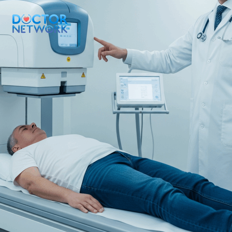
The MRI examination takes place in a specialized suite containing the large, cylindrical scanner and associated monitoring equipment. You’ll lie on a movable table that slides into the scanner opening, with positioning depending on the body area being examined.
Absolute stillness is crucial throughout the scanning process to prevent motion artifacts that could compromise image quality. The technologist may ask you to hold your breath for short periods during certain sequences, particularly for abdominal or cardiac imaging.
MRI scanners produce loud tapping, knocking, and buzzing sounds during image acquisition. You’ll receive ear protection (earplugs or headphones) to reduce noise exposure, and many facilities offer music options to make the experience more comfortable.
Communication with the MRI technologist remains possible throughout the examination via intercom system. The technologist can see and hear you at all times, and you can request to stop the scan if necessary. A friend or family member may be permitted in the scanning room if they meet safety requirements.
Contrast injection typically occurs partway through the examination, administered through the previously established IV line. You may experience a cool sensation, metallic taste, or mild warmth during injection, but these sensations are normal and temporary.
Scan Duration and Expectations
MRI examinations typically require 15-90 minutes depending on the body area being studied and the number of sequences needed. Contrast-enhanced studies generally take longer than non-contrast examinations due to additional imaging sequences performed after contrast administration.
Typical examination durations:
- Brain MRI with contrast: 30-45 minutes
- Abdominal MRI with contrast: 45-60 minutes
- Cardiac MRI with contrast: 60-90 minutes
- Spine MRI with contrast: 30-60 minutes
- Pelvic MRI with contrast: 45-75 minutes
Post-Examination Care and Recovery
Immediate Post-Scan Activities
Most MRI examinations are outpatient procedures allowing immediate return to normal activities unless sedation was administered. Patients who received only contrast enhancement without sedation can typically resume work, driving, and regular daily activities immediately after the scan.
Following sedation, patients require extended observation and must have a responsible adult provide transportation home. Recovery from conscious sedation typically takes 2-4 hours, during which alertness and coordination gradually return to normal levels.
Contrast Agent Elimination and Hydration
Drinking plenty of water after contrast-enhanced MRI helps accelerate gadolinium elimination through the kidneys. While not medically necessary for patients with normal kidney function, increased fluid intake can help reduce any minor side effects and promote faster contrast clearance.
The contrast agent is typically eliminated from the body within 24-48 hours through normal kidney filtration. Patients with impaired kidney function may have prolonged elimination times and require additional monitoring.
Monitoring for Delayed Reactions
While immediate severe reactions to gadolinium are rare, patients should remain alert for delayed allergic reactions that can occur hours after contrast administration. Symptoms requiring immediate medical attention include difficulty breathing, widespread rash or hives, severe nausea or vomiting, or significant swelling of face, lips, or throat.
Signs of delayed contrast reactions:
- Skin rash, itching, or hives developing 1-7 days post-contrast
- Nausea, vomiting, or abdominal cramping
- Joint pain or swelling
- Headache or dizziness
- Unusual fatigue or weakness
Contact your healthcare provider if you experience any concerning symptoms after contrast administration. While serious delayed reactions are uncommon, prompt medical evaluation ensures appropriate treatment if complications develop.
Results Timeline and Follow-up Planning
MRI results are not immediately available following your examination. A specialized radiologist must analyze the images and prepare a detailed report describing the findings and their clinical significance. This interpretation process typically requires 1-2 weeks for routine examinations, though urgent cases may receive expedited reporting.
Your referring physician will receive the radiology report and contact you to discuss the results and any necessary follow-up care. Avoid attempting to interpret the images yourself, as proper analysis requires specialized training and knowledge of normal anatomical variations.
Understanding Contrast-Related Risks and Safety Measures
Common Side Effects and Management
Gadolinium-based contrast agents demonstrate excellent safety profiles with adverse reactions occurring in less than 1% of patients. Most side effects are mild and temporary, resolving without specific treatment within a few hours of contrast administration.
Mild, common side effects include:
- Temporary nausea or mild stomach upset
- Cool sensation or metallic taste during injection
- Minor headache or dizziness
- Slight pain or warmth at the injection site
These minor reactions rarely require treatment and typically resolve spontaneously. Staying well-hydrated and avoiding sudden position changes can help minimize these mild effects.
Serious Complications and Risk Factors
While severe contrast reactions are rare, certain patient populations face increased risks requiring careful evaluation before contrast administration. Patients with severely impaired kidney function (eGFR < 30 mL/min/1.73m²) may develop nephrogenic systemic fibrosis, a serious condition affecting skin and internal organs.
Previous allergic reactions to gadolinium contrast, while uncommon, increase the risk of repeat reactions. Patients with known contrast allergies may require premedication with antihistamines and corticosteroids before repeat contrast exposure.
FDA Safety Guidelines and Regulatory Oversight
The Food and Drug Administration continues to monitor gadolinium contrast safety through post-market surveillance programs and periodic safety reviews. Current FDA guidance supports the continued use of approved gadolinium agents for appropriate clinical indications while acknowledging small risks in susceptible populations.
Recent FDA communications have highlighted concerns about gadolinium retention in brain tissue following repeated contrast exposures, though no adverse clinical effects have been definitively established. This ongoing research emphasizes the importance of using contrast agents only when medically justified and choosing the safest available formulations.
5 common questions about
1. Why do you have to fast before an MRI with contrast?
Fasting is necessary before an MRI with contrast to ensure clear and accurate imaging. When your stomach is empty, the contrast agent can be absorbed and distributed more effectively without interference from food or digestive activity. This improves the quality of the images and helps radiologists detect abnormalities more precisely. Additionally, fasting reduces the risk of nausea and vomiting caused by the contrast injection, which could be dangerous during the scan when you are lying down.
2. How long should you fast before an MRI with contrast?
The typical fasting period recommended is about 4 to 6 hours before the MRI scan. During this time, you should avoid eating any food or drinking anything except small sips of water. This duration helps ensure the stomach is empty and the contrast agent is not diluted or affected by recent food intake, leading to optimal image quality and patient safety.
3. What are the risks if you do not fast before the MRI with contrast?
Not fasting can increase the risk of vomiting and aspiration, where stomach contents might be inhaled into the lungs, causing respiratory complications. Food in the digestive tract can also create artifacts or distortions in the MRI images, making it harder to interpret the results accurately. Furthermore, the contrast agent may not distribute evenly, reducing the diagnostic effectiveness of the scan.
4. Can you drink water during the fasting period before the MRI with contrast?
Yes, drinking small amounts of water is usually allowed during the fasting period. Water does not interfere with the absorption or distribution of the contrast agent and helps prevent dehydration. However, other beverages and food should be avoided to maintain an empty stomach for the best imaging results.
5. Are there exceptions or special considerations for fasting before MRI with contrast?
Yes, patients with certain medical conditions such as diabetes, kidney or liver problems, or gastrointestinal issues may need tailored fasting instructions. In some cases, alternative preparation methods or modified fasting protocols may be used under medical supervision to balance safety and diagnostic needs. Always consult your healthcare provider for personalized guidance before the scan
Conclusion
Fasting before contrast-enhanced MRI serves dual critical purposes: ensuring patient safety by preventing potentially life-threatening aspiration complications and optimizing diagnostic image quality through motion artifact reduction. These requirements represent evidence-based medical protocols developed to maximize both safety and diagnostic accuracy during these sophisticated imaging procedures.
The specific fasting duration and restrictions vary based on the examination type, patient characteristics, and institutional protocols, ranging from 4-8 hours for most procedures. Cardiac MRI examinations require unique considerations including extended caffeine avoidance, while procedures requiring sedation mandate complete NPO status for extended periods.
Successful MRI preparation requires clear communication with your healthcare team, careful attention to fasting guidelines, appropriate medication management, and thorough safety screening. Patients with diabetes, kidney disease, or other medical conditions need individualized preparation strategies to ensure both safety and diagnostic success.
Always consult directly with your physician or the radiology department for precise instructions tailored to your specific examination and medical history. Following these evidence-based guidelines ensures the safest possible experience while obtaining the highest quality diagnostic images needed for accurate medical diagnosis and treatment planning. Your cooperation with these important safety protocols contributes significantly to the success of your medical care and helps maintain the excellent safety record of modern contrast-enhanced MRI imaging.
References
Nausea and Vomiting as a Side Effect of Contrast Media:
Gadolinium-based contrast agents, while generally safe, can sometimes cause side effects, including nausea and, less commonly, vomiting.
Evidence:
Source: American College of Radiology (ACR) Manual on Contrast Media. This is a regularly updated, authoritative guide for radiologists and healthcare professionals.
Authors: The manual is a committee document, with numerous expert contributors from the ACR Committee on Drugs and Contrast Media.
Relevant Information: The ACR Manual (e.g., Version 2023 or similar recent versions) lists nausea and vomiting as known “mild” acute adverse reactions to GBCAs. While the incidence is relatively low (nausea is more common than vomiting), the potential exists.
Quote/Paraphrase: “Mild reactions [to GBCAs] include nausea, vomiting, warmth or coldness, headache, dizziness, itching, flushing… Nausea and vomiting are among the most common physiologic reactions.” (This is a general statement often found in such manuals).
Specific Study Example (Illustrative – exact incidence rates vary by specific GBCA and study):
Hunt, C. H., Hartman, R. P., & Hesley, G. K. (2009). Frequency and severity of adverse effects of iodinated and gadolinium contrast materials: a review of 456,930 doses. American Journal of Roentgenology, 193(4), 1124-1127.
Finding: This study, while also covering iodinated contrast, provides data on adverse reactions to GBCAs. It would show that nausea and vomiting occur, albeit typically at low rates (e.g., nausea around 1-2%, vomiting less than 1%). The key is that they can occur.
Risk of Aspiration:
If a patient vomits while lying flat on their back (supine position) in the MRI scanner, there is a significant risk that the vomitus could be aspirated into the lungs.
The MRI procedure can be lengthy, and the patient is often in a confined space, making it difficult to quickly sit up or turn if they feel nauseous.
Evidence: This is a general medical principle rather than a specific MRI study. The risk of aspiration during procedures where a patient is supine and might vomit is well-established in medicine and anesthesiology.
Source: Standard medical textbooks on patient safety, anesthesia, or emergency medicine.
Authors: Various.
Relevant Principle: Fasting reduces gastric volume and acidity, thereby decreasing the quantity and potential harm of aspirate if vomiting occurs. This is a standard precaution before many medical procedures, especially those involving sedation or where the patient is supine.
Fasting Guidelines are Precautionary:
Although severe reactions leading to vomiting are uncommon with modern GBCAs, the policy of fasting is a conservative, safety-first approach.
The potential consequences of aspiration (e.g., chemical pneumonitis, bacterial pneumonia) are serious enough to warrant this precaution.
Source: Hospital and imaging department protocols worldwide.
Rationale: These protocols are developed based on overarching patient safety guidelines, recommendations from professional bodies like the ACR, and an understanding of potential adverse events.
What if Sedation is Used?
If a patient requires sedation or general anesthesia for the MRI (e.g., due to claustrophobia, pain, or inability to remain still, common in pediatric MRIs), fasting becomes even more critical.
Sedatives can depress protective airway reflexes (like the gag reflex), significantly increasing the risk of aspiration if vomiting occurs.
Evidence: Anesthesiology guidelines.
Source: American Society of Anesthesiologists (ASA) Task Force on Preoperative Fasting.
Authors: ASA Committee.
Relevant Guideline: “Practice Guidelines for Preoperative Fasting and the Use of Pharmacologic Agents to Reduce the Risk of Pulmonary Aspiration: Application to Healthy Patients Undergoing Elective Procedures.” (Published periodically in Anesthesiology). These guidelines clearly state fasting requirements before procedures involving sedation or anesthesia to minimize aspiration risk.
Kiểm Duyệt Nội Dung
More than 10 years of marketing communications experience in the medical and health field.
Successfully deployed marketing communication activities, content development and social networking channels for hospital partners, clinics, doctors and medical professionals across the country.
More than 6 years of experience in organizing and producing leading prestigious medical programs in Vietnam, in collaboration with Ho Chi Minh City Television (HTV). Typical programs include Nhật Ký Blouse Trắng, Bác Sĩ Nói Gì, Alo Bác Sĩ Nghe, Nhật Ký Hạnh Phúc, Vui Khỏe Cùng Con, Bác Sỹ Mẹ, v.v.
Comprehensive cooperation with hundreds of hospitals and clinics, thousands of doctors and medical experts to join hands in building a medical content and service platform on the Doctor Network application.