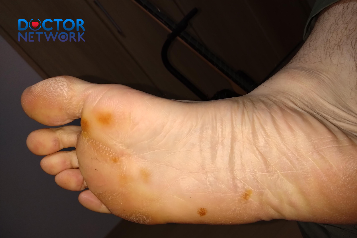
Dark or brown spots appearing on the sole of your foot represent a spectrum of conditions ranging from completely harmless pigmentation to potentially life-threatening melanoma. These plantar lesions, skin discolorations, and foot blemishes affect millions of people annually, yet many individuals remain unaware of when these dermal manifestations require immediate medical attention versus simple observation. Understanding the distinction between benign pigmented lesions and malignant skin cancer on your feet could literally save your life, as foot melanoma often goes undiagnosed until advanced stages due to its inconspicuous location.
Navigating Dark Spots on Bottom of Feet – This comprehensive guide will examine the critical differences between harmless and concerning foot spots, teach you the essential ABCDE self-examination technique for identifying suspicious lesions, explore common causes including acral melanoma and tinea nigra fungal infections, and explain when urgent medical evaluation becomes necessary. We’ll also cover the diagnostic procedures your healthcare provider will perform and treatment approaches for various conditions affecting the plantar surface of your feet.
Not All Spots Are Created Equal: Benign vs. Concerning

The appearance of dark spots on feet varies dramatically in size, shape, color intensity, and underlying pathology. Benign lesions typically maintain consistent characteristics over time, while malignant transformations often exhibit irregular features that evolve progressively. This fundamental distinction between non-cancerous and potentially cancerous skin abnormalities forms the cornerstone of proper foot dermatology assessment.
Healthcare professionals utilize specific diagnostic criteria to differentiate between harmless pigmented nevi (moles), post-inflammatory hyperpigmentation, and aggressive skin cancers like acral lentiginous melanoma. Visual examination alone cannot provide definitive diagnosis, making professional medical evaluation essential for any concerning plantar lesion. Dermatoscopic analysis, biopsy procedures, and histopathological examination remain the gold standard for accurate diagnosis of suspicious foot spots.
The critical nature of this distinction cannot be overstated—while benign conditions may require no treatment beyond monitoring, malignant lesions demand immediate surgical intervention and oncological management to prevent metastatic spread and improve survival outcomes.
Identifying Suspicious Spots: Applying the ABCDE Rule on the Foot
The ABCDE rule serves as your primary screening tool for detecting potentially malignant skin lesions, including those located on the challenging-to-examine plantar surface. Each letter represents a crucial warning sign that warrants immediate dermatological consultation and possible biopsy evaluation.
The ABCDE Criteria for Foot Examination
Asymmetry occurs when one half of the spot fails to match the other half in shape, color distribution, or border characteristics. Benign moles typically display symmetrical patterns, while melanomas often exhibit irregular, lopsided appearances that suggest malignant cellular growth patterns.
Border Irregularity manifests as ragged, notched, scalloped, or blurred edges surrounding the pigmented lesion. Normal moles maintain smooth, well-defined borders, whereas cancerous spots frequently display jagged perimeters that reflect uncontrolled cellular proliferation.
Color Variation within a single spot indicates potential malignancy, particularly when multiple shades of brown, black, tan, red, white, or blue appear together. Benign lesions usually maintain uniform coloration, while melanomas often develop heterogeneous pigmentation patterns.
Diameter measurements exceeding 6 millimeters (approximately pencil eraser size) raise concern for malignant transformation, though smaller melanomas can certainly occur and should not be dismissed based solely on size criteria.
Evolution represents the most critical warning sign—any changes in size, shape, color, texture, or symptomatology over time demands immediate medical evaluation, as benign lesions rarely undergo significant transformation.
Self-Check Guide for Plantar Examination
Regular foot inspection requires systematic methodology to ensure comprehensive coverage of all plantar surfaces, including difficult-to-visualize areas between toes and along the heel margins.
Monthly Examination Protocol:
- Use a handheld mirror or request assistance from a family member for complete visualization
- Examine each foot in bright lighting conditions, preferably natural daylight
- Document any new spots with photographs for comparison tracking
- Note changes in existing lesions using the ABCDE criteria
- Pay special attention to areas of previous trauma or chronic friction
High-Risk Locations:
- Weight-bearing surfaces (heel, ball of foot, big toe)
- Areas between toes and nail beds
- Previous injury sites or chronic pressure points
- Regions with poor circulation or diabetic complications
Common Causes of Dark Spots on the Bottom of Feet
Multiple pathological processes can produce dark pigmentation on plantar surfaces, ranging from infectious organisms to malignant cellular transformations. Understanding these diverse etiologies enables appropriate risk stratification and treatment planning.
Melanoma on the Sole: Why It’s High-Risk (Acral Melanoma)
Acral lentiginous melanoma represents the most dangerous cause of dark spots on feet, accounting for the majority of melanomas in individuals with darker skin tones and carrying significantly higher mortality rates due to delayed diagnosis. This aggressive skin cancer typically develops on palms, soles, and subungual (under nail) locations where sun exposure plays minimal causative roles.
The delayed recognition of acral melanoma stems from its atypical location and appearance compared to sun-induced melanomas on other body regions. Patients and healthcare providers often dismiss plantar lesions as calluses, bruises, or fungal infections, leading to diagnostic delays that allow metastatic progression.
Characteristic Features of Acral Melanoma:
- Irregular, asymmetrical borders with color variation
- Progressive enlargement over weeks to months
- Development in previously normal skin areas
- Potential for ulceration or bleeding in advanced stages
- Absence of typical melanoma risk factors (sun exposure, fair skin)
The prognosis for acral melanoma correlates directly with thickness at diagnosis, emphasizing the critical importance of early detection and prompt surgical management.
Tinea Nigra: The Fungal Imposter
Tinea nigra presents as a superficial fungal infection caused by Hortaea werneckii, creating dark brown or black patches that frequently mimic melanoma on clinical examination. This tropical and subtropical mycosis affects palms and soles, producing asymptomatic hyperpigmented lesions that can cause significant diagnostic confusion.
The organism Hortaea werneckii thrives in warm, humid environments and typically produces single, non-scaly patches with well-defined borders. Unlike bacterial or inflammatory skin conditions, tinea nigra rarely causes itching, pain, or secondary symptoms, making it easily overlooked or misdiagnosed.
Clinical Characteristics:
- Single, sharply demarcated dark patch
- Non-inflammatory appearance without scaling
- Asymptomatic presentation (no itching or discomfort)
- Gradual enlargement over months
- Positive response to antifungal therapy
Diagnostic confirmation requires skin scraping with potassium hydroxide (KOH) preparation or fungal culture, revealing characteristic branching hyphae and yeast forms under microscopic examination.
Common Benign Causes: Bruises, Moles, and Pigmentation
Traumatic Bruising frequently produces dark discoloration on plantar surfaces following injury, repetitive pressure, or microtrauma from ill-fitting footwear. These subungual or subcutaneous hemorrhages typically display characteristic color evolution from dark red-purple to brown-yellow as hemoglobin breakdown products are reabsorbed.
Plantar bruises often result from:
- Athletic activities with repetitive foot impact
- Foreign object puncture wounds
- Chronic pressure from tight footwear
- Workplace injuries in industrial settings
Benign Melanocytic Nevi (Moles) can develop on any skin surface, including plantar regions, typically appearing as uniform brown or black spots with regular borders and consistent coloration. These common pigmented lesions rarely undergo malignant transformation but require monitoring for changes suggestive of dysplastic evolution.
Post-inflammatory Hyperpigmentation develops following skin trauma, infection, or inflammatory conditions, leaving darkened areas that gradually fade over months to years. Common triggers include:
- Resolved bacterial or fungal infections
- Healing surgical wounds or lacerations
- Chronic dermatitis or eczematous conditions
- Chemical burns from topical medications
Diabetes and Foot Spots: The Connection
Diabetic patients face elevated risks for various foot complications, including specific dermatological manifestations that can appear as dark spots or patches on plantar surfaces. Poor circulation, neuropathy, and impaired immune function create conditions favoring skin changes and delayed healing responses.
Diabetic Dermopathy presents as small, brown, scaly patches primarily affecting the anterior shins but potentially involving foot surfaces in individuals with long-standing diabetes mellitus. These lesions result from microangiopathy and typically appear as multiple, round-to-oval hyperpigmented areas with slight scale or atrophy.
Table 1: Diabetic Foot Complications and Associated Dark Spots
| Condition | Appearance | Location | Risk Factors |
|---|---|---|---|
| Diabetic Dermopathy | Small brown patches with scale | Shins, occasionally feet | Poor glucose control, duration >10 years |
| Acanthosis Nigricans | Velvety hyperpigmentation | Skin folds, occasionally plantar | Insulin resistance, obesity |
| Necrobiosis Lipoidica | Yellow-brown plaques | Pretibial area, rarely feet | Type 1 diabetes, female gender |
| Chronic Ulceration | Dark, callused margins | Pressure points on feet | Neuropathy, poor circulation |
Other Potential Causes
Vascular Conditions can produce dark spots through various mechanisms including small vessel thrombosis, chronic venous insufficiency, or arterial compromise leading to tissue hypoxia and pigmentation changes.
Friction Calluses may darken over time due to chronic trauma, embedded debris, or secondary bacterial colonization, particularly in individuals who walk barefoot frequently or wear inadequate footwear protection.
Foreign Body Reactions occasionally present as dark spots when metallic objects, glass fragments, or organic materials become embedded in plantar tissues, triggering inflammatory responses and secondary pigmentation.
When to See a Doctor: Recognizing Warning Signs

Immediate medical evaluation becomes essential when dark spots on feet display characteristics suggesting malignant potential or represent symptoms of serious underlying conditions. Delaying professional assessment of suspicious lesions can result in diagnostic delays that significantly impact treatment outcomes and survival rates.
Critical Warning Signs Requiring Urgent Evaluation
New Spot Development in previously normal skin areas warrants dermatological consultation, particularly in individuals over age 50 or those with family histories of melanoma or other skin cancers.
Progressive Changes in existing spots using ABCDE criteria indicate potential malignant transformation and demand immediate biopsy evaluation to rule out melanoma or other aggressive skin cancers.
Symptomatic Lesions causing itching, bleeding, pain, or ulceration suggest inflammatory processes or malignant changes that require prompt medical intervention and possible surgical management.
Irregular Features including asymmetrical borders, color variation, or diameter exceeding 6 millimeters raise significant concern for acral melanoma and necessitate urgent dermatological referral.
Risk Stratification Checklist
Utilize this systematic approach to determine urgency of medical consultation:
High Priority (Urgent – Within 1-2 weeks):
- Any spot meeting ABCDE criteria
- New lesions in patients >50 years old
- Bleeding, ulcerated, or painful spots
- Family history of melanoma plus new spots
Moderate Priority (Within 1 month):
- Stable spots in high-risk individuals
- Diabetic patients with new foot lesions
- Immunocompromised patients with skin changes
- Occupational exposure to carcinogens
Low Priority (Routine monitoring):
- Stable benign-appearing moles
- Known post-traumatic pigmentation
- Chronic, unchanged callused areas
Medical Diagnosis Explained: What Your Doctor Will Do
Healthcare providers follow systematic diagnostic protocols to accurately identify the underlying cause of plantar dark spots, utilizing clinical examination, specialized equipment, and laboratory testing when appropriate.
Initial Clinical Assessment begins with comprehensive visual examination of the concerning lesion and surrounding skin areas, documenting size, shape, color characteristics, and border irregularities using standardized photography and measurement techniques.
Dermatoscopic Evaluation employs specialized handheld microscopes to examine surface patterns, vascular structures, and pigmentation distribution not visible to naked eye examination, significantly improving diagnostic accuracy for melanoma detection.
Medical History Documentation includes family cancer history, previous skin cancer diagnoses, medication usage, occupational exposures, and symptom timeline to establish risk factors and narrow differential diagnoses.
Diagnostic Procedures by Suspected Condition
For Suspected Melanoma:
- Full-body skin examination for additional lesions
- Dermatoscopic pattern analysis
- Excisional biopsy with adequate margins
- Sentinel lymph node evaluation if indicated
- Staging studies for confirmed malignancy
For Suspected Tinea Nigra:
- Potassium hydroxide (KOH) preparation of skin scrapings
- Fungal culture on Sabouraud dextrose agar
- Wood’s lamp examination (may show fluorescence)
- Response to topical antifungal therapy
For Diabetic Complications:
- Comprehensive foot examination with monofilament testing
- Vascular assessment with ankle-brachial index
- Glycemic control evaluation
- Wound culture if secondary infection suspected
Table 2: Diagnostic Tests for Common Foot Spot Conditions
| Suspected Diagnosis | Primary Test | Secondary Tests | Typical Results |
|---|---|---|---|
| Melanoma | Excisional biopsy | Dermatoscopy, imaging | Atypical melanocytes, mitotic activity |
| Tinea Nigra | KOH preparation | Fungal culture | Branching hyphae, yeast forms |
| Benign Nevus | Clinical exam | Dermatoscopy | Regular pattern, uniform color |
| Diabetic Dermopathy | Clinical diagnosis | Glucose testing | Correlation with diabetes control |
| Traumatic Bruise | History, exam | None usually needed | Consistent with trauma timeline |
Treatment Approaches (Brief Overview)
Treatment strategies depend entirely on accurate diagnosis of the underlying condition causing plantar dark spots, ranging from simple observation to urgent surgical intervention with adjuvant therapy.
Antifungal Therapy for confirmed tinea nigra typically involves topical agents such as ketoconazole, terbinafine, or ciclopirox applied twice daily for 2-4 weeks until complete resolution occurs. Systemic antifungal medications rarely become necessary except in extensive or resistant cases.
Surgical Management for melanoma requires wide local excision with appropriate margins based on tumor thickness, potentially followed by sentinel lymph node biopsy and adjuvant immunotherapy or targeted therapy for high-risk cases.
Conservative Monitoring applies to benign lesions including stable moles, post-inflammatory hyperpigmentation, and traumatic bruising, with periodic reassessment to ensure no malignant transformation occurs.
Treatment Success Factors
Patient Compliance with prescribed therapies and follow-up appointments directly correlates with treatment outcomes, particularly for chronic conditions requiring prolonged management.
Early Intervention provides optimal results for all conditions, from simple fungal infections to aggressive melanomas, emphasizing the importance of prompt medical evaluation for concerning lesions.
Multidisciplinary Care may become necessary for complex cases involving diabetes management, wound care, infectious disease consultation, or oncological treatment planning.
Table 3: Treatment Timeline and Expected Outcomes
| Condition | Treatment Duration | Expected Improvement | Follow-up Requirements |
|---|---|---|---|
| Tinea Nigra | 2-4 weeks topical antifungal | Complete resolution | None after cure |
| Benign Nevus | Observation only | Stable appearance | Annual skin checks |
| Melanoma (early) | Surgery + possible adjuvant | Depends on staging | Lifelong monitoring |
| Diabetic Dermopathy | Glucose control | Gradual fading | Ongoing diabetes care |
| Post-inflammatory Hyperpigmentation | 6-12 months | Slow lightening | Monitor for recurrence |
Conclusion: Prioritizing Your Foot Health
Dark spots appearing on the bottom of your feet encompass a broad spectrum of conditions, from completely harmless post-traumatic pigmentation to life-threatening acral melanoma requiring immediate surgical intervention. The key to optimal outcomes lies in understanding when these plantar lesions warrant professional medical evaluation versus simple home monitoring using established clinical criteria.
Regular self-examination using the ABCDE methodology enables early detection of suspicious changes that might otherwise go unnoticed until advanced stages develop. Monthly foot inspections should become routine healthcare maintenance, particularly for individuals with diabetes, family cancer histories, or occupational risk factor exposures.
Remember that only qualified healthcare providers can provide definitive diagnoses through appropriate clinical examination, dermatoscopic evaluation, and histopathological analysis when indicated. Never attempt to self-diagnose concerning skin lesions, as delayed treatment of malignant conditions can result in preventable complications and reduced survival rates.
Prioritize your foot health by maintaining awareness of normal versus abnormal skin changes, seeking prompt medical attention for suspicious lesions, and following recommended screening guidelines for your individual risk profile. Early detection and appropriate treatment of foot spot conditions can prevent serious complications while ensuring optimal long-term outcomes for your overall health and wellbeing.
5 common questions
1. What causes dark spots on the bottom of feet?
Dark spots on the bottom of feet can be caused by several factors including:
Hyperpigmentation due to excess melanin from sun exposure or aging
Friction and pressure leading to calluses or bruises
Fungal infections such as tinea nigra or athlete’s foot
Skin conditions like stasis dermatitis or diabetic dermopathy
More serious causes like plantar warts (caused by HPV) or malignant melanoma (a type of skin cancer)1457.
2. Are dark spots on the feet dangerous?
Most dark spots are harmless and related to skin thickening, bruising, or minor infections. However, some dark spots can be serious, especially if they are new, changing in size, shape, or color, or accompanied by pain or swelling. Melanomas on the feet can appear as dark spots and require early medical evaluation to prevent severe outcomes.
3. How can I tell if a dark spot on my foot is melanoma?
Signs that a dark spot might be melanoma include:
Irregular shape or border
Multiple colors within the spot
Changes in size, shape, or color over time
Itching, bleeding, or pain
Size larger than a quarter of an inch
If you notice these signs, see a dermatologist promptly for diagnosis.
4. What treatments are available for dark spots on the bottom of feet?
Treatment depends on the cause:
For plantar warts (dark spots caused by HPV), over-the-counter salicylic acid treatments or medical removal (cryotherapy, surgery) are options
Fungal infections can be treated with antifungal creams
Calluses and friction-related spots may improve with protective footwear and moisturizing
Melanomas require surgical removal and oncological management
For hyperpigmentation, sun protection and topical lightening agents may help.
5. How can I prevent dark spots on my feet?
Preventive measures include:
Protecting feet from excessive sun exposure using sunscreen or protective clothing
Wearing shoes or socks in public wet areas to avoid HPV and fungal infections
Inspecting feet regularly for any new or changing spots
Managing underlying health conditions like diabetes to improve circulation and skin health
Avoiding prolonged friction or pressure on feet by wearing well-fitting shoes.
References
Acral Lentiginous Melanoma (ALM)
Description: This is a rare but aggressive type of skin cancer that can occur on the palms, soles, or under the nails. It’s more common in people with darker skin tones. Early detection is critical.
Evidence/Study: “Acral Lentiginous Melanoma: A Clinicopathologic and Outcome Study of 126 Cases.”
Authors: Kuchelmeister, C., Schaumburg-Lever, G., Garbe, C.
Source: British Journal of Dermatology, 2000, 143(2), 275-280.
Key finding (generalized from this and similar studies): ALM often presents as a dark brown to black, irregularly shaped macule or patch that may grow slowly. It can be misdiagnosed, leading to delays in treatment.
Link (to abstract/related info): While the direct full text might be behind a paywall, you can often find abstracts on PubMed. A general link to information on ALM from a reputable source:
Skin Cancer Foundation: https://www.skincancer.org/skin-cancer-information/melanoma/acral-lentiginous-melanoma/
American Academy of Dermatology (AAD) on melanoma: https://www.aad.org/public/diseases/skin-cancer/types/common/melanoma (Search for “acral” on their site).
Study: “Acral Lentiginous Melanoma: Incidence and Survival Patterns in the United States, 1986-2005.”
Authors: Bradford PT, Goldstein AM, McMaster ML, Tucker MA.
Source: Archives of Dermatology (now JAMA Dermatology), 2009;145(4):427–434.
Key finding: This study analyzed incidence and survival rates, highlighting the importance of understanding this subtype of melanoma.
Link: https://jamanetwork.com/journals/jamadermatology/fullarticle/712060
Junctional Nevi (Moles)
Description: These are common, benign (non-cancerous) moles that are flat and typically occur where the epidermis (outer skin layer) meets the dermis (inner skin layer). They can be present on the soles.
Evidence/Study: “Plantar Warts Versus Plantar Moles (Acral Nevi).”
Authors: (This is often covered in dermatology textbooks and review articles rather than specific pinpoint studies unless focusing on dermoscopy or differentiation from melanoma). For general information from a reliable source:
Source: DermNet NZ (a highly respected dermatological resource).
Key finding (general dermatological knowledge): Junctional nevi on the soles are common and usually harmless, but any change in size, shape, color, or symptoms (like itching or bleeding) warrants investigation.
Link: https://dermnetnz.org/topics/melanocytic-naevus-of-sole
Tinea Nigra
Description: A superficial fungal infection that causes light brown to black, flat patches, typically on the palms or soles. It’s painless and not usually itchy.
Evidence/Study: “Tinea Nigra: An Updated Review.”
Authors: Xavier, M. H., & de Oliveira, J. C.
Source: Mycopathologia, 2020, 185(4), 585-599.
Key finding: This review discusses the epidemiology, clinical presentation, diagnosis (often by KOH smear or culture), and treatment of Tinea Nigra. It can mimic melanoma, so correct diagnosis is important.
Link (to abstract): https://link.springer.com/article/10.1007/s11046-020-00441-x
Another good general resource: StatPearls (NCBI) – “Tinea Nigra”
Authors: Many contributing authors, updated regularly. (Example: Gupta G, Mahajan K.)
Source: StatPearls Publishing; 2023 Jan-.
Post-Inflammatory Hyperpigmentation (PIH)
Description: Darkening of the skin that occurs after an injury or inflammation (like a blister, cut, burn, or eczema) has healed.
Evidence/Study: “Postinflammatory Hyperpigmentation: A Review of the Epidemiology, Clinical Features, and Treatment Options in Skin of Color.”
Authors: Davis, E. C., & Callender, V. D.
Source: The Journal of Clinical and Aesthetic Dermatology, 2010, 3(7), 20–31.
Key finding: PIH is common, especially in individuals with darker skin tones. It results from increased melanin production or deposition after skin inflammation.
Talon Noir (Black Heel or Palm)
Description: This appears as a group of small, dark dots or a larger dark patch on the heel or palm, caused by minor bleeding (hemorrhage) into the skin due to shearing stress from activities like running or sports. It’s harmless.
Evidence/Study: “Black Heel and Palm (Talon Noir).”
Authors: (Often described in sports medicine and dermatology case reports and reviews).
Source: For example, a chapter in a dermatology textbook or a review article on sports-related dermatoses. DermNet NZ provides a good overview.
Key finding: Caused by trauma leading to intraepidermal hemorrhage. Paring the skin (scraping the top layer) will reveal pinpoint bleeding spots if it’s talon noir, distinguishing it from melanoma.
Stains or Foreign Bodies
Description: Dyes from socks or shoes, dirt, or small splinters can cause dark spots.
Evidence/Study: This is generally based on clinical observation rather than formal studies. A doctor can often determine this by examining the spot and taking a patient history.
No specific scientific study for “stains,” but dermatologists are trained to recognize these.
Kiểm Duyệt Nội Dung
More than 10 years of marketing communications experience in the medical and health field.
Successfully deployed marketing communication activities, content development and social networking channels for hospital partners, clinics, doctors and medical professionals across the country.
More than 6 years of experience in organizing and producing leading prestigious medical programs in Vietnam, in collaboration with Ho Chi Minh City Television (HTV). Typical programs include Nhật Ký Blouse Trắng, Bác Sĩ Nói Gì, Alo Bác Sĩ Nghe, Nhật Ký Hạnh Phúc, Vui Khỏe Cùng Con, Bác Sỹ Mẹ, v.v.
Comprehensive cooperation with hundreds of hospitals and clinics, thousands of doctors and medical experts to join hands in building a medical content and service platform on the Doctor Network application.


